 Movie
Movie Controller

[English] 日本語
 Yorodumi
- PDB-6zum: Crystal structure of dimethylated RSL-N23H (RSL-B3) in complex wi... -
+ Open data

- Basic information
| Entry | Database: PDB / ID: 6zum | ||||||||||||
|---|---|---|---|---|---|---|---|---|---|---|---|---|---|
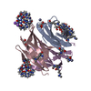
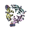
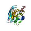
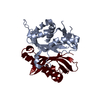
| Title | Crystal structure of dimethylated RSL-N23H (RSL-B3) in complex with cucurbit[7]uril and zinc | ||||||||||||
 Components Components | Fucose-binding lectin protein | ||||||||||||
 Keywords Keywords | SUGAR BINDING PROTEIN / cucurbituril / molecular glue / crystal engineering / dimethyllysine / zinc / b-propeller | ||||||||||||
| Function / homology | Fucose-specific lectin / Fungal fucose-specific lectin / carbohydrate binding / metal ion binding / ACETATE ION / beta-D-fructopyranose / cucurbit[7]uril / Fucose-binding lectin protein Function and homology information Function and homology information | ||||||||||||
| Biological species |  Ralstonia solanacearum (bacteria) Ralstonia solanacearum (bacteria) | ||||||||||||
| Method |  X-RAY DIFFRACTION / SYNCHROTRON / MOLECULAR REPLACEMENT / Resolution: 1.589 Å X-RAY DIFFRACTION / SYNCHROTRON / MOLECULAR REPLACEMENT / Resolution: 1.589 Å | ||||||||||||
 Authors Authors | Guagnini, F. / Engilberge, S. / Flood, R.J. / Ramberg, K.O. / Crowley, P.B. | ||||||||||||
| Funding support |  Ireland, 3items Ireland, 3items
| ||||||||||||
 Citation Citation | Journal: Cryst.Growth Des. / Year: 2020 Title: Metal-Mediated Protein-Cucurbituril Crystalline Architectures Authors: Guagnini, F. / Engilberge, S. / Flood, R.J. / Ramberg, K.O. / Crowley, P.B. | ||||||||||||
| History |
|
- Structure visualization
| Structure viewer | Molecule: MolmilJmol/JSmol |
|---|
- Downloads & links
-Download
| PDBx/mmCIF format | 6zum.cif.gz | 63.4 KB | Display | PDBx/mmCIF format |
|---|---|---|---|---|
| PDB format | pdb6zum.ent.gz | 44.7 KB | Display | PDB format |
| PDBx/mmJSON format | 6zum.json.gz | Tree view | PDBx/mmJSON format | |
| Others |  Other downloads Other downloads |
-Validation report
| Arichive directory | https://data.pdbj.org/pub/pdb/validation_reports/zu/6zumftp://data.pdbj.org/pub/pdb/validation_reports/zu/6zum | HTTPS FTP |
|---|
-Related structure data
| Related structure data |  6zukC  6zulC  6f7wS S: Starting model for refinement C: citing same article ( |
|---|---|
| Similar structure data |


-Links
 PDBj
PDBj
- Assembly
| Deposited unit | 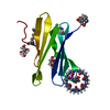
| ||||||||||||
|---|---|---|---|---|---|---|---|---|---|---|---|---|---|
| 1 | 
| ||||||||||||
| Unit cell |
| ||||||||||||
| Components on special symmetry positions |
|
-Components
-Protein / Sugars , 2 types, 3 molecules A
| #1: Protein | Mass: 9866.798 Da / Num. of mol.: 1 Source method: isolated from a genetically manipulated source Source: (gene. exp.) Ralstonia solanacearum (bacteria)Gene: E7Z57_08365, RSP795_21825, RSP822_19650, RUN39_v1_50103 Production host: |
|---|---|
| #2: Sugar |  Type: D-saccharide, beta linking / Mass: 180.156 Da / Num. of mol.: 2 / Source method: obtained synthetically / Formula: C6H12O6 Type: D-saccharide, beta linking / Mass: 180.156 Da / Num. of mol.: 2 / Source method: obtained synthetically / Formula: C6H12O6 |
-Non-polymers , 5 types, 62 molecules 


| #3: Chemical | ChemComp-ACT /  Mass: 59.044 Da / Num. of mol.: 1 / Source method: obtained synthetically / Formula: C2H3O2 Mass: 59.044 Da / Num. of mol.: 1 / Source method: obtained synthetically / Formula: C2H3O2 | ||||||
|---|---|---|---|---|---|---|---|
| #4: Chemical |  Mass: 65.409 Da / Num. of mol.: 2 / Source method: obtained synthetically / Formula: Zn / Feature type: SUBJECT OF INVESTIGATION Mass: 65.409 Da / Num. of mol.: 2 / Source method: obtained synthetically / Formula: Zn / Feature type: SUBJECT OF INVESTIGATION#5: Chemical | ChemComp-QQ7 / |  Mass: 1162.962 Da / Num. of mol.: 1 / Source method: obtained synthetically / Formula: C42H42N28O14 / Feature type: SUBJECT OF INVESTIGATION Mass: 1162.962 Da / Num. of mol.: 1 / Source method: obtained synthetically / Formula: C42H42N28O14 / Feature type: SUBJECT OF INVESTIGATION#6: Chemical |  Mass: 22.990 Da / Num. of mol.: 2 / Source method: obtained synthetically / Formula: Na Mass: 22.990 Da / Num. of mol.: 2 / Source method: obtained synthetically / Formula: Na#7: Water | ChemComp-HOH / | Mass: 18.015 Da / Num. of mol.: 56 / Source method: isolated from a natural source / Formula: H2O |
-Details
| Has ligand of interest | Y |
|---|
-Experimental details
-Experiment
| Experiment | Method: X-RAY DIFFRACTION / Number of used crystals: 1 |
|---|
- Sample preparation
| Crystal | Density Matthews: 2.33 Å3/Da / Density % sol: 47.21 % |
|---|---|
| Crystal grow | Temperature: 293 K / Method: vapor diffusion, sitting drop Details: 10% PEG 8000, 0.1 M TRIS-HCl pH 7.0, 0.2 M magnesium chloride |
-Data collection
| Diffraction | Mean temperature: 100 K / Serial crystal experiment: N |
|---|---|
| Diffraction source | Source: SYNCHROTRON / Site: SOLEIL  / Beamline: PROXIMA 2 / Wavelength: 0.98013 Å / Beamline: PROXIMA 2 / Wavelength: 0.98013 Å |
| Detector | Type: DECTRIS EIGER X 9M / Detector: PIXEL / Date: Feb 1, 2019 |
| Radiation | Protocol: SINGLE WAVELENGTH / Monochromatic (M) / Laue (L): M / Scattering type: x-ray |
| Radiation wavelength | Wavelength: 0.98013 Å / Relative weight: 1 |
| Reflection | Resolution: 1.589→34.159 Å / Num. obs: 13378 / % possible obs: 99.7 % / Redundancy: 9 % / CC1/2: 0.996 / Rmerge(I) obs: 0.091 / Rpim(I) all: 0.03 / Rrim(I) all: 0.096 / Net I/σ(I): 14.3 |
| Reflection shell | Resolution: 1.589→1.617 Å / Rmerge(I) obs: 0.692 / Mean I/σ(I) obs: 2.1 / Num. unique obs: 646 / CC1/2: 0.775 / Rpim(I) all: 0.332 / Rrim(I) all: 0.771 |
- Processing
| Software |
| ||||||||||||||||||||||||||||||||||||||||||
|---|---|---|---|---|---|---|---|---|---|---|---|---|---|---|---|---|---|---|---|---|---|---|---|---|---|---|---|---|---|---|---|---|---|---|---|---|---|---|---|---|---|---|---|
| Refinement | Method to determine structure: MOLECULAR REPLACEMENT Starting model: 6F7W polyalanine Resolution: 1.589→34.159 Å / SU ML: 0.18 / Cross valid method: THROUGHOUT / σ(F): 2.03 / Phase error: 25.75 / Stereochemistry target values: ML
| ||||||||||||||||||||||||||||||||||||||||||
| Solvent computation | Shrinkage radii: 0.9 Å / VDW probe radii: 1.11 Å / Solvent model: FLAT BULK SOLVENT MODEL | ||||||||||||||||||||||||||||||||||||||||||
| Displacement parameters | Biso max: 96.71 Å2 / Biso mean: 43.2139 Å2 / Biso min: 16.94 Å2 | ||||||||||||||||||||||||||||||||||||||||||
| Refinement step | Cycle: final / Resolution: 1.589→34.159 Å
| ||||||||||||||||||||||||||||||||||||||||||
| Refine LS restraints |
| ||||||||||||||||||||||||||||||||||||||||||
| LS refinement shell | Refine-ID: X-RAY DIFFRACTION / Rfactor Rfree error: 0
| ||||||||||||||||||||||||||||||||||||||||||
| Refinement TLS params. | Method: refined / Origin x: 6.809 Å / Origin y: 9.9386 Å / Origin z: 0.4441 Å
| ||||||||||||||||||||||||||||||||||||||||||
| Refinement TLS group |
|
