 Movie
Movie Controller

[English] 日本語
 Yorodumi
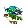
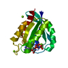
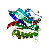
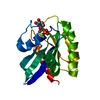
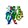
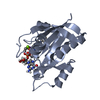
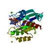
- PDB-6zli: CRYSTAL STRUCTURE OF KRAS-G12D IN COMPLEX WITH COMPOUND 13 AND GCP -
+ Open data

- Basic information
| Entry | Database: PDB / ID: 6zli | ||||||
|---|---|---|---|---|---|---|---|
| Title | CRYSTAL STRUCTURE OF KRAS-G12D IN COMPLEX WITH COMPOUND 13 AND GCP | ||||||
 Components Components | GTPase KRas | ||||||
 Keywords Keywords | HYDROLASE / GTPase | ||||||
| Function / homology |  Function and homology information Function and homology informationresponse to mineralocorticoid / GMP binding / forebrain astrocyte development / LRR domain binding / regulation of synaptic transmission, GABAergic / negative regulation of epithelial cell differentiation / response to isolation stress / response to gravity / epithelial tube branching involved in lung morphogenesis / type I pneumocyte differentiation ...response to mineralocorticoid / GMP binding / forebrain astrocyte development / LRR domain binding / regulation of synaptic transmission, GABAergic / negative regulation of epithelial cell differentiation / response to isolation stress / response to gravity / epithelial tube branching involved in lung morphogenesis / type I pneumocyte differentiation / Rac protein signal transduction / myoblast proliferation / Signaling by RAS GAP mutants / Signaling by RAS GTPase mutants / Activation of RAS in B cells / RAS signaling downstream of NF1 loss-of-function variants / RUNX3 regulates p14-ARF / positive regulation of glial cell proliferation / skeletal muscle cell differentiation / SOS-mediated signalling / Activated NTRK3 signals through RAS / Activated NTRK2 signals through RAS / cardiac muscle cell proliferation / SHC1 events in ERBB4 signaling / Signalling to RAS / SHC-related events triggered by IGF1R / Activated NTRK2 signals through FRS2 and FRS3 / Estrogen-stimulated signaling through PRKCZ / positive regulation of Rac protein signal transduction / SHC-mediated cascade:FGFR3 / glial cell proliferation / MET activates RAS signaling / SHC-mediated cascade:FGFR2 / SHC-mediated cascade:FGFR4 / Signaling by PDGFRA transmembrane, juxtamembrane and kinase domain mutants / Signaling by PDGFRA extracellular domain mutants / PTK6 Regulates RHO GTPases, RAS GTPase and MAP kinases / Erythropoietin activates RAS / SHC-mediated cascade:FGFR1 / Signaling by FGFR4 in disease / Signaling by CSF3 (G-CSF) / FRS-mediated FGFR3 signaling / Signaling by FLT3 ITD and TKD mutants / FRS-mediated FGFR2 signaling / FRS-mediated FGFR4 signaling / p38MAPK events / striated muscle cell differentiation / FRS-mediated FGFR1 signaling / Signaling by FGFR3 in disease / Tie2 Signaling / protein-membrane adaptor activity / Signaling by FGFR2 in disease / Signaling by FLT3 fusion proteins / GRB2 events in EGFR signaling / SHC1 events in EGFR signaling / FLT3 Signaling / Signaling by FGFR1 in disease / EGFR Transactivation by Gastrin / NCAM signaling for neurite out-growth / CD209 (DC-SIGN) signaling / Downstream signal transduction / GRB2 events in ERBB2 signaling / homeostasis of number of cells within a tissue / Insulin receptor signalling cascade / SHC1 events in ERBB2 signaling / Constitutive Signaling by Overexpressed ERBB2 / response to glucocorticoid / Signaling by phosphorylated juxtamembrane, extracellular and kinase domain KIT mutants / Ras activation upon Ca2+ influx through NMDA receptor / VEGFR2 mediated cell proliferation / small monomeric GTPase / FCERI mediated MAPK activation / liver development / female pregnancy / Signaling by ERBB2 TMD/JMD mutants / Signaling by SCF-KIT / RAF activation / Constitutive Signaling by EGFRvIII / Signaling by high-kinase activity BRAF mutants / Signaling by ERBB2 ECD mutants / MAP2K and MAPK activation / Signaling by ERBB2 KD Mutants / visual learning / regulation of long-term neuronal synaptic plasticity / cytoplasmic side of plasma membrane / cytokine-mediated signaling pathway / Signaling by CSF1 (M-CSF) in myeloid cells / Signaling by RAF1 mutants / Signaling by moderate kinase activity BRAF mutants / Paradoxical activation of RAF signaling by kinase inactive BRAF / Signaling downstream of RAS mutants / Negative regulation of MAPK pathway / Regulation of RAS by GAPs / RAS processing / positive regulation of cellular senescence / Signaling by BRAF and RAF1 fusions / DAP12 signaling / GDP binding / MAPK cascade / Constitutive Signaling by Ligand-Responsive EGFR Cancer Variants Similarity search - Function | ||||||
| Biological species |  Homo sapiens (human) Homo sapiens (human) | ||||||
| Method |  X-RAY DIFFRACTION / MOLECULAR REPLACEMENT / Resolution: 1.73 Å X-RAY DIFFRACTION / MOLECULAR REPLACEMENT / Resolution: 1.73 Å | ||||||
 Authors Authors | Kessler, D. / Fischer, G. / Boettcher, J. | ||||||
 Citation Citation | Journal: Future Med Chem / Year: 2020 Title: Drugging all RAS isoforms with one pocket. Authors: Kessler, D. / Bergner, A. / Bottcher, J. / Fischer, G. / Dobel, S. / Hinkel, M. / Mullauer, B. / Weiss-Puxbaum, A. / McConnell, D.B. | ||||||
| History |
|
- Structure visualization
| Structure viewer | Molecule: MolmilJmol/JSmol |
|---|
- Downloads & links
-Download
| PDBx/mmCIF format | 6zli.cif.gz | 154.4 KB | Display | PDBx/mmCIF format |
|---|---|---|---|---|
| PDB format | pdb6zli.ent.gz | 121 KB | Display | PDB format |
| PDBx/mmJSON format | 6zli.json.gz | Tree view | PDBx/mmJSON format | |
| Others |  Other downloads Other downloads |
-Validation report
| Arichive directory | https://data.pdbj.org/pub/pdb/validation_reports/zl/6zliftp://data.pdbj.org/pub/pdb/validation_reports/zl/6zli | HTTPS FTP |
|---|
-Related structure data
| Related structure data |  6zioC  6zirC  6zizC 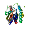 6zj0C 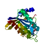 6zl3C  6zl5C  6gj8S S: Starting model for refinement C: citing same article ( |
|---|---|
| Similar structure data |


-Links
 PDBj
PDBj


- Assembly
| Deposited unit | 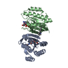
| ||||||||
|---|---|---|---|---|---|---|---|---|---|
| 1 | 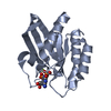
| ||||||||
| 2 | 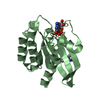
| ||||||||
| Unit cell |
|
-Components
| #1: Protein | Mass: 19386.848 Da / Num. of mol.: 2 Source method: isolated from a genetically manipulated source Source: (gene. exp.) Homo sapiens (human) / Gene: KRAS, KRAS2, RASK2 / Production host:  #2: Chemical |   Mass: 521.208 Da / Num. of mol.: 2 / Source method: obtained synthetically / Formula: C11H18N5O13P3 / Comment: GMP-PCP, energy-carrying molecule analogue*YM Mass: 521.208 Da / Num. of mol.: 2 / Source method: obtained synthetically / Formula: C11H18N5O13P3 / Comment: GMP-PCP, energy-carrying molecule analogue*YM#3: Chemical |   Mass: 24.305 Da / Num. of mol.: 2 / Source method: obtained synthetically / Formula: Mg Mass: 24.305 Da / Num. of mol.: 2 / Source method: obtained synthetically / Formula: Mg#4: Chemical | ChemComp-QME / |   Mass: 200.280 Da / Num. of mol.: 1 / Source method: isolated from a natural source / Formula: C13H16N2 / Feature type: SUBJECT OF INVESTIGATION Mass: 200.280 Da / Num. of mol.: 1 / Source method: isolated from a natural source / Formula: C13H16N2 / Feature type: SUBJECT OF INVESTIGATION#5: Water | ChemComp-HOH / |  Mass: 18.015 Da / Num. of mol.: 261 / Source method: isolated from a natural source / Formula: H2O Mass: 18.015 Da / Num. of mol.: 261 / Source method: isolated from a natural source / Formula: H2OHas ligand of interest | Y | |
|---|
-Experimental details
-Experiment
| Experiment | Method: X-RAY DIFFRACTION / Number of used crystals: 1 |
|---|
- Sample preparation
| Crystal | Density Matthews: 2.16 Å3/Da / Density % sol: 43.01 % |
|---|---|
| Crystal grow | Temperature: 278 K / Method: vapor diffusion / Details: 51.8% MPD 50mM MES PH= 6.4 |
-Data collection
| Diffraction | Mean temperature: 100 K / Serial crystal experiment: N |
|---|---|
| Diffraction source | Source: SEALED TUBE / Type: RIGAKU MICROMAX-003 / Wavelength: 1.5418 Å |
| Detector | Type: RIGAKU SATURN 944 / Detector: CCD / Date: Aug 5, 2015 |
| Radiation | Protocol: SINGLE WAVELENGTH / Monochromatic (M) / Laue (L): M / Scattering type: x-ray |
| Radiation wavelength | Wavelength: 1.5418 Å / Relative weight: 1 |
| Reflection | Resolution: 1.73→42.7915 Å / Num. obs: 23941 / % possible obs: 80.7 % / Redundancy: 3.1 % / Rmerge(I) obs: 0.051 / Rsym value: 0.051 / Net I/σ(I): 15.6 |
| Reflection shell | Resolution: 1.747→1.862 Å / Mean I/σ(I) obs: 1.8 / Num. unique obs: 1197 / CC1/2: 0.62 |
- Processing
| Software |
| |||||||||||||||||||||||||||||||||||||||||||||||||||||||||||||||||||||||||||
|---|---|---|---|---|---|---|---|---|---|---|---|---|---|---|---|---|---|---|---|---|---|---|---|---|---|---|---|---|---|---|---|---|---|---|---|---|---|---|---|---|---|---|---|---|---|---|---|---|---|---|---|---|---|---|---|---|---|---|---|---|---|---|---|---|---|---|---|---|---|---|---|---|---|---|---|---|
| Refinement | Method to determine structure: MOLECULAR REPLACEMENT Starting model: 6GJ8 Resolution: 1.73→42.8 Å / Cor.coef. Fo:Fc: 0.942 / Cor.coef. Fo:Fc free: 0.917 / SU R Cruickshank DPI: 0.175 / Cross valid method: THROUGHOUT / SU R Blow DPI: 0.184 / SU Rfree Blow DPI: 0.154 / SU Rfree Cruickshank DPI: 0.152
| |||||||||||||||||||||||||||||||||||||||||||||||||||||||||||||||||||||||||||
| Displacement parameters | Biso mean: 17.67 Å2
| |||||||||||||||||||||||||||||||||||||||||||||||||||||||||||||||||||||||||||
| Refine analyze | Luzzati coordinate error obs: 0.23 Å | |||||||||||||||||||||||||||||||||||||||||||||||||||||||||||||||||||||||||||
| Refinement step | Cycle: LAST / Resolution: 1.73→42.8 Å
| |||||||||||||||||||||||||||||||||||||||||||||||||||||||||||||||||||||||||||
| Refine LS restraints |
| |||||||||||||||||||||||||||||||||||||||||||||||||||||||||||||||||||||||||||
| LS refinement shell | Resolution: 1.73→1.81 Å /
| |||||||||||||||||||||||||||||||||||||||||||||||||||||||||||||||||||||||||||
| Refinement TLS params. | Refine-ID: X-RAY DIFFRACTION
| |||||||||||||||||||||||||||||||||||||||||||||||||||||||||||||||||||||||||||
| Refinement TLS group |
|
