 Movie
Movie Controller

[English] 日本語
 Yorodumi
- PDB-6zgq: AceL NrdHF class 3 split intein GSH linked splice inactive varian... -
+ Open data

- Basic information
| Entry | Database: PDB / ID: 6zgq | ||||||
|---|---|---|---|---|---|---|---|
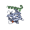
| Title | AceL NrdHF class 3 split intein GSH linked splice inactive variant - C124A, N146A | ||||||
 Components Components | AceL NrdHF-1-1 Intein | ||||||
 Keywords Keywords | SPLICING | ||||||
| Function / homology | Endonuclease - Pi-scei; Chain A, domain 1 / Hedgehog/Intein (Hint) domain / Beta Complex / Mainly Beta / IODIDE ION Function and homology information Function and homology information | ||||||
| Biological species | metagenome (others) | ||||||
| Method |  X-RAY DIFFRACTION / SYNCHROTRON / SAD / Resolution: 1.9 Å X-RAY DIFFRACTION / SYNCHROTRON / SAD / Resolution: 1.9 Å | ||||||
 Authors Authors | Hoffmann, S. / Mootz, H.D. / Kuemmel, D. / Singh, R. | ||||||
| Funding support | 1items
| ||||||
 Citation Citation | Journal: Chembiochem / Year: 2021 Title: Biochemical and Structural Characterization of an Unusual and Naturally Split Class 3 Intein. Authors: Hoffmann, S. / Terhorst, T.M.E. / Singh, R.K. / Kummel, D. / Pietrokovski, S. / Mootz, H.D. | ||||||
| History |
|
- Structure visualization
| Structure viewer | Molecule: MolmilJmol/JSmol |
|---|
- Downloads & links
-Download
| PDBx/mmCIF format | 6zgq.cif.gz | 143.3 KB | Display | PDBx/mmCIF format |
|---|---|---|---|---|
| PDB format | pdb6zgq.ent.gz | 113.5 KB | Display | PDB format |
| PDBx/mmJSON format | 6zgq.json.gz | Tree view | PDBx/mmJSON format | |
| Others |  Other downloads Other downloads |
-Validation report
| Arichive directory | https://data.pdbj.org/pub/pdb/validation_reports/zg/6zgqftp://data.pdbj.org/pub/pdb/validation_reports/zg/6zgq | HTTPS FTP |
|---|
-Related structure data
| Similar structure data |
|---|


-Links
 PDBj
PDBj- Assembly
| Deposited unit | 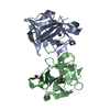
| ||||||||
|---|---|---|---|---|---|---|---|---|---|
| 1 | 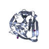
| ||||||||
| 2 | 
| ||||||||
| Unit cell |
|
-Components
| #1: Protein | Mass: 18591.914 Da / Num. of mol.: 2 Source method: isolated from a genetically manipulated source Source: (gene. exp.) metagenome (others) / Production host:  #2: Chemical | ChemComp-IOD /   Mass: 126.904 Da / Num. of mol.: 7 / Source method: obtained synthetically / Formula: I Mass: 126.904 Da / Num. of mol.: 7 / Source method: obtained synthetically / Formula: I#3: Water | ChemComp-HOH / |  Mass: 18.015 Da / Num. of mol.: 192 / Source method: isolated from a natural source / Formula: H2O Mass: 18.015 Da / Num. of mol.: 192 / Source method: isolated from a natural source / Formula: H2OHas ligand of interest | N | |
|---|
-Experimental details
-Experiment
| Experiment | Method: X-RAY DIFFRACTION / Number of used crystals: 1 |
|---|
- Sample preparation
| Crystal | Density Matthews: 1.97 Å3/Da / Density % sol: 37.71 % |
|---|---|
| Crystal grow | Temperature: 305 K / Method: vapor diffusion, hanging drop / Details: PEG3350 25 %, Glycerol 20 %, 0.1 M MIB |
-Data collection
| Diffraction | Mean temperature: 100 K / Serial crystal experiment: N | ||||||||||||||||||||||||||||||
|---|---|---|---|---|---|---|---|---|---|---|---|---|---|---|---|---|---|---|---|---|---|---|---|---|---|---|---|---|---|---|---|
| Diffraction source | Source: SYNCHROTRON / Site: BESSY  / Beamline: 14.2 / Wavelength: 1.5498 Å / Beamline: 14.2 / Wavelength: 1.5498 Å | ||||||||||||||||||||||||||||||
| Detector | Type: DECTRIS PILATUS3 2M / Detector: PIXEL / Date: Jan 27, 2020 | ||||||||||||||||||||||||||||||
| Radiation | Protocol: SINGLE WAVELENGTH / Monochromatic (M) / Laue (L): M / Scattering type: x-ray | ||||||||||||||||||||||||||||||
| Radiation wavelength | Wavelength: 1.5498 Å / Relative weight: 1 | ||||||||||||||||||||||||||||||
| Reflection | Resolution: 1.9→41.96 Å / Num. obs: 20988 / % possible obs: 93.6 % / Redundancy: 3.4 % / CC1/2: 0.993 / Rmerge(I) obs: 0.09 / Rpim(I) all: 0.058 / Rrim(I) all: 0.107 / Net I/σ(I): 7.1 / Num. measured all: 72086 | ||||||||||||||||||||||||||||||
| Reflection shell | Diffraction-ID: 1
|
- Processing
| Software |
| |||||||||||||||||||||||||||||||||||||||||||||||||||||||||||||||||||||||||||
|---|---|---|---|---|---|---|---|---|---|---|---|---|---|---|---|---|---|---|---|---|---|---|---|---|---|---|---|---|---|---|---|---|---|---|---|---|---|---|---|---|---|---|---|---|---|---|---|---|---|---|---|---|---|---|---|---|---|---|---|---|---|---|---|---|---|---|---|---|---|---|---|---|---|---|---|---|
| Refinement | Method to determine structure: SAD / Resolution: 1.9→41.96 Å / Cor.coef. Fo:Fc: 0.922 / Cor.coef. Fo:Fc free: 0.91 / SU B: 13.883 / SU ML: 0.173 / Cross valid method: THROUGHOUT / σ(F): 0 / ESU R Free: 0.185 / Stereochemistry target values: MAXIMUM LIKELIHOOD Details: HYDROGENS HAVE BEEN ADDED IN THE RIDING POSITIONS U VALUES : REFINED INDIVIDUALLY
| |||||||||||||||||||||||||||||||||||||||||||||||||||||||||||||||||||||||||||
| Solvent computation | Ion probe radii: 0.8 Å / Shrinkage radii: 0.8 Å / VDW probe radii: 1.2 Å / Solvent model: MASK | |||||||||||||||||||||||||||||||||||||||||||||||||||||||||||||||||||||||||||
| Displacement parameters | Biso max: 291.7 Å2 / Biso mean: 15.512 Å2 / Biso min: 4.86 Å2
| |||||||||||||||||||||||||||||||||||||||||||||||||||||||||||||||||||||||||||
| Refinement step | Cycle: final / Resolution: 1.9→41.96 Å
| |||||||||||||||||||||||||||||||||||||||||||||||||||||||||||||||||||||||||||
| Refine LS restraints |
| |||||||||||||||||||||||||||||||||||||||||||||||||||||||||||||||||||||||||||
| LS refinement shell | Resolution: 1.9→1.949 Å / Rfactor Rfree error: 0 / Total num. of bins used: 20
|
