 Movie
Movie Controller

[English] 日本語
 Yorodumi
- PDB-6z61: Crystal structure of NAD kinase 1 from Listeria monocytogenes in ... -
+ Open data

- Basic information
| Entry | Database: PDB / ID: 6z61 | ||||||
|---|---|---|---|---|---|---|---|
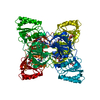
| Title | Crystal structure of NAD kinase 1 from Listeria monocytogenes in complex with a di-adenosine derivative | ||||||
 Components Components | NAD kinase 1 | ||||||
 Keywords Keywords | TRANSFERASE / tetrameric NAD-kinase | ||||||
| Function / homology |  Function and homology information Function and homology informationNAD+ kinase / NAD+ kinase activity / NADP+ biosynthetic process / NAD+ metabolic process / NAD binding / ATP binding / metal ion binding / cytoplasm Similarity search - Function | ||||||
| Biological species |  Listeria monocytogenes EGD-e (bacteria) Listeria monocytogenes EGD-e (bacteria) | ||||||
| Method |  X-RAY DIFFRACTION / SYNCHROTRON / MOLECULAR REPLACEMENT / Resolution: 2.47 Å X-RAY DIFFRACTION / SYNCHROTRON / MOLECULAR REPLACEMENT / Resolution: 2.47 Å | ||||||
 Authors Authors | Gelin, M. / Labesse, G. | ||||||
 Citation Citation | Journal: Molecules / Year: 2020 Title: New Chemical Probe Targeting Bacterial NAD Kinase. Authors: Clement, D.A. / Leseigneur, C. / Gelin, M. / Coelho, D. / Huteau, V. / Lionne, C. / Labesse, G. / Dussurget, O. / Pochet, S. | ||||||
| History |
|
- Structure visualization
| Structure viewer | Molecule: MolmilJmol/JSmol |
|---|
- Downloads & links
-Download
| PDBx/mmCIF format | 6z61.cif.gz | 122.1 KB | Display | PDBx/mmCIF format |
|---|---|---|---|---|
| PDB format | pdb6z61.ent.gz | 93.8 KB | Display | PDB format |
| PDBx/mmJSON format | 6z61.json.gz | Tree view | PDBx/mmJSON format | |
| Others |  Other downloads Other downloads |
-Validation report
| Arichive directory | https://data.pdbj.org/pub/pdb/validation_reports/z6/6z61ftp://data.pdbj.org/pub/pdb/validation_reports/z6/6z61 | HTTPS FTP |
|---|
-Related structure data
| Related structure data |  6z64C  6z65C  6rg9S S: Starting model for refinement C: citing same article ( |
|---|---|
| Similar structure data |


-Links
 PDBj
PDBj- Assembly
| Deposited unit | 
| ||||||||||
|---|---|---|---|---|---|---|---|---|---|---|---|
| 1 | 
| ||||||||||
| Unit cell |
| ||||||||||
| Components on special symmetry positions |
|
-Components
| #1: Protein | Mass: 31045.279 Da / Num. of mol.: 1 Source method: isolated from a genetically manipulated source Source: (gene. exp.) Listeria monocytogenes EGD-e (bacteria)Gene: nadK1, lmo0968 / Production host: |
|---|---|
| #2: Chemical | ChemComp-CIT /   Mass: 192.124 Da / Num. of mol.: 1 / Source method: obtained synthetically / Formula: C6H8O7 Mass: 192.124 Da / Num. of mol.: 1 / Source method: obtained synthetically / Formula: C6H8O7 |
| #3: Chemical | ChemComp-Q9H / (  Mass: 613.583 Da / Num. of mol.: 1 / Source method: obtained synthetically / Formula: C25H31N11O8 / Feature type: SUBJECT OF INVESTIGATION Mass: 613.583 Da / Num. of mol.: 1 / Source method: obtained synthetically / Formula: C25H31N11O8 / Feature type: SUBJECT OF INVESTIGATION |
| #4: Water | ChemComp-HOH /  Mass: 18.015 Da / Num. of mol.: 15 / Source method: isolated from a natural source / Formula: H2O Mass: 18.015 Da / Num. of mol.: 15 / Source method: isolated from a natural source / Formula: H2O |
| Has ligand of interest | Y |
-Experimental details
-Experiment
| Experiment | Method: X-RAY DIFFRACTION / Number of used crystals: 1 |
|---|
- Sample preparation
| Crystal | Density Matthews: 2.25 Å3/Da / Density % sol: 45.22 % |
|---|---|
| Crystal grow | Temperature: 291.15 K / Method: vapor diffusion, hanging drop Details: 30 mM NaBr, 220 mM Kcitrate, glycerol 6%, 15-16% w/v PEG400 PH range: 4.8 - 5.1 |
-Data collection
| Diffraction | Mean temperature: 100 K / Serial crystal experiment: N | ||||||||||||||||||||||||||||||
|---|---|---|---|---|---|---|---|---|---|---|---|---|---|---|---|---|---|---|---|---|---|---|---|---|---|---|---|---|---|---|---|
| Diffraction source | Source: SYNCHROTRON / Site: ESRF  / Beamline: MASSIF-1 / Wavelength: 0.966 Å / Beamline: MASSIF-1 / Wavelength: 0.966 Å | ||||||||||||||||||||||||||||||
| Detector | Type: DECTRIS PILATUS3 2M / Detector: PIXEL / Date: Jul 16, 2018 | ||||||||||||||||||||||||||||||
| Radiation | Protocol: SINGLE WAVELENGTH / Monochromatic (M) / Laue (L): M / Scattering type: x-ray | ||||||||||||||||||||||||||||||
| Radiation wavelength | Wavelength: 0.966 Å / Relative weight: 1 | ||||||||||||||||||||||||||||||
| Reflection | Resolution: 2.241→59.07 Å / Num. obs: 11262 / % possible obs: 89 % / Redundancy: 9.9 % / CC1/2: 0.999 / Rmerge(I) obs: 0.06 / Rpim(I) all: 0.019 / Rrim(I) all: 0.063 / Net I/σ(I): 22.1 | ||||||||||||||||||||||||||||||
| Reflection shell | Diffraction-ID: 1
|
- Processing
| Software |
| ||||||||||||||||||||||||||||||||||||||||||||||||||||||||||||||||||||||||||||||||||||||||||||||||||||
|---|---|---|---|---|---|---|---|---|---|---|---|---|---|---|---|---|---|---|---|---|---|---|---|---|---|---|---|---|---|---|---|---|---|---|---|---|---|---|---|---|---|---|---|---|---|---|---|---|---|---|---|---|---|---|---|---|---|---|---|---|---|---|---|---|---|---|---|---|---|---|---|---|---|---|---|---|---|---|---|---|---|---|---|---|---|---|---|---|---|---|---|---|---|---|---|---|---|---|---|---|---|
| Refinement | Method to determine structure: MOLECULAR REPLACEMENT Starting model: 6RG9 Resolution: 2.47→59.07 Å / SU ML: 0.27 / Cross valid method: THROUGHOUT / σ(F): 1.34 / Phase error: 31.85 / Stereochemistry target values: ML
| ||||||||||||||||||||||||||||||||||||||||||||||||||||||||||||||||||||||||||||||||||||||||||||||||||||
| Solvent computation | Shrinkage radii: 0.9 Å / VDW probe radii: 1.11 Å / Solvent model: FLAT BULK SOLVENT MODEL | ||||||||||||||||||||||||||||||||||||||||||||||||||||||||||||||||||||||||||||||||||||||||||||||||||||
| Displacement parameters | Biso max: 115.28 Å2 / Biso mean: 65.3517 Å2 / Biso min: 32.8 Å2 | ||||||||||||||||||||||||||||||||||||||||||||||||||||||||||||||||||||||||||||||||||||||||||||||||||||
| Refinement step | Cycle: final / Resolution: 2.47→59.07 Å
| ||||||||||||||||||||||||||||||||||||||||||||||||||||||||||||||||||||||||||||||||||||||||||||||||||||
| LS refinement shell | Refine-ID: X-RAY DIFFRACTION / Rfactor Rfree error: 0 / Total num. of bins used: 3
| ||||||||||||||||||||||||||||||||||||||||||||||||||||||||||||||||||||||||||||||||||||||||||||||||||||
| Refinement TLS params. | Method: refined / Refine-ID: X-RAY DIFFRACTION
| ||||||||||||||||||||||||||||||||||||||||||||||||||||||||||||||||||||||||||||||||||||||||||||||||||||
| Refinement TLS group |
|
