 Movie
Movie Controller

[English] 日本語
 Yorodumi
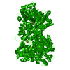
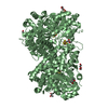
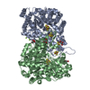
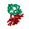
- PDB-6yu9: CO-dehydrogenase homodimer from Clostridium autoethanogenum at 1.... -
+ Open data

- Basic information
| Entry | Database: PDB / ID: 6yu9 | |||||||||
|---|---|---|---|---|---|---|---|---|---|---|
| Title | CO-dehydrogenase homodimer from Clostridium autoethanogenum at 1.90-A resolution | |||||||||
 Components Components | Carbon-monoxide dehydrogenase (Acceptor),Carbon-monoxide dehydrogenase (Acceptor) | |||||||||
 Keywords Keywords | OXIDOREDUCTASE / C1-Metabolism / Carbon monoxide / metallo-containing protein / Acetogenic bacteria / Acetogenesis / CO-dehydrogenase/Acetyl-CoA synthase / X-ray crystal structure / Gas channeling / Waste-gas conversion | |||||||||
| Function / homology |  Function and homology information Function and homology informationcarbon-monoxide dehydrogenase (acceptor) / : / nickel cation binding / generation of precursor metabolites and energy / 4 iron, 4 sulfur cluster binding Similarity search - Function | |||||||||
| Biological species |  Clostridium autoethanogenum DSM 10061 (bacteria) Clostridium autoethanogenum DSM 10061 (bacteria) | |||||||||
| Method |  X-RAY DIFFRACTION / SYNCHROTRON / MOLECULAR REPLACEMENT / Resolution: 1.904 Å X-RAY DIFFRACTION / SYNCHROTRON / MOLECULAR REPLACEMENT / Resolution: 1.904 Å | |||||||||
 Authors Authors | Wagner, T. / Lemaire, O.N. | |||||||||
| Funding support |  Germany, 2items Germany, 2items
| |||||||||
 Citation Citation | Journal: Biochim Biophys Acta Bioenerg / Year: 2020 Title: Gas channel rerouting in a primordial enzyme: Structural insights of the carbon-monoxide dehydrogenase/acetyl-CoA synthase complex from the acetogen Clostridium autoethanogenum. Authors: Lemaire, O.N. / Wagner, T. | |||||||||
| History |
|
- Structure visualization
| Structure viewer | Molecule: MolmilJmol/JSmol |
|---|
- Downloads & links
-Download
| PDBx/mmCIF format | 6yu9.cif.gz | 946 KB | Display | PDBx/mmCIF format |
|---|---|---|---|---|
| PDB format | pdb6yu9.ent.gz | 786 KB | Display | PDB format |
| PDBx/mmJSON format | 6yu9.json.gz | Tree view | PDBx/mmJSON format | |
| Others |  Other downloads Other downloads |
-Validation report
| Arichive directory | https://data.pdbj.org/pub/pdb/validation_reports/yu/6yu9ftp://data.pdbj.org/pub/pdb/validation_reports/yu/6yu9 | HTTPS FTP |
|---|
-Related structure data
| Related structure data | 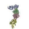 6yttSC 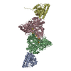 6yuaC S: Starting model for refinement C: citing same article ( |
|---|---|
| Similar structure data |


-Links
 PDBj
PDBj- Assembly
| Deposited unit | 
| ||||||||
|---|---|---|---|---|---|---|---|---|---|
| 1 | 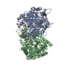
| ||||||||
| 2 | 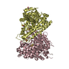
| ||||||||
| Unit cell |
|
-Components
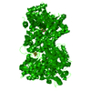
-Protein , 1 types, 4 molecules ABCD
| #1: Protein | Mass: 67977.531 Da / Num. of mol.: 4 / Mutation: Wild-type / Source method: isolated from a natural source Details: A stop codon is present in the gene and the CODH seems to be split in two peptides. However, experimental evidence proved that a stop codon read-through occurs leading to a cysteine (C401). ...Details: A stop codon is present in the gene and the CODH seems to be split in two peptides. However, experimental evidence proved that a stop codon read-through occurs leading to a cysteine (C401).,A stop codon is present in the gene and the CODH seems to be split in two peptides. However, experimental evidence proved that a stop codon read-through occurs leading to a cysteine (C401). Source: (natural) Clostridium autoethanogenum DSM 10061 (bacteria)Cell line: / / Organ: / / Plasmid details: / / Variant: / / Tissue: / References: UniProt: U5RTE2, carbon-monoxide dehydrogenase (acceptor) |
|---|
-Non-polymers , 6 types, 1125 molecules 

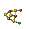


| #2: Chemical | ChemComp-ACT /  Mass: 59.044 Da / Num. of mol.: 5 / Source method: obtained synthetically / Formula: C2H3O2 Mass: 59.044 Da / Num. of mol.: 5 / Source method: obtained synthetically / Formula: C2H3O2#3: Chemical | ChemComp-SF4 /  Mass: 351.640 Da / Num. of mol.: 6 / Source method: obtained synthetically / Formula: Fe4S4 / Feature type: SUBJECT OF INVESTIGATION Mass: 351.640 Da / Num. of mol.: 6 / Source method: obtained synthetically / Formula: Fe4S4 / Feature type: SUBJECT OF INVESTIGATION#4: Chemical | ChemComp-XCC /  Mass: 410.333 Da / Num. of mol.: 4 / Source method: obtained synthetically / Formula: Fe4NiS4 / Feature type: SUBJECT OF INVESTIGATION Mass: 410.333 Da / Num. of mol.: 4 / Source method: obtained synthetically / Formula: Fe4NiS4 / Feature type: SUBJECT OF INVESTIGATION#5: Chemical |  Mass: 62.068 Da / Num. of mol.: 2 / Source method: obtained synthetically / Formula: C2H6O2 Mass: 62.068 Da / Num. of mol.: 2 / Source method: obtained synthetically / Formula: C2H6O2#6: Chemical | ChemComp-CL / |  Mass: 35.453 Da / Num. of mol.: 1 / Source method: obtained synthetically / Formula: Cl Mass: 35.453 Da / Num. of mol.: 1 / Source method: obtained synthetically / Formula: Cl#7: Water | ChemComp-HOH / | Mass: 18.015 Da / Num. of mol.: 1107 / Source method: isolated from a natural source / Formula: H2O |
|---|
-Details
| Has ligand of interest | Y |
|---|
-Experimental details
-Experiment
| Experiment | Method: X-RAY DIFFRACTION / Number of used crystals: 1 |
|---|
- Sample preparation
| Crystal | Density Matthews: 2.56 Å3/Da / Density % sol: 52.05 % / Description: Brownish thick quadratic plates |
|---|---|
| Crystal grow | Temperature: 291.15 K / Method: vapor diffusion, sitting drop / pH: 7.6 Details: Crystallization was performed in an anoxic chamber (N2/H2, 95:5 %) with anaerobic solutions. Crystals were obtained by initial screening at 291.15 K using the sitting drop method on a 96- ...Details: Crystallization was performed in an anoxic chamber (N2/H2, 95:5 %) with anaerobic solutions. Crystals were obtained by initial screening at 291.15 K using the sitting drop method on a 96-well MRC 2 Crystallization Plates in polystyrene (SWISSCI). The crystallization reservoir contained 90 uL of mother liquor, crystallization drop contained a mixture of 0.6 uL protein and 0.6 uL precipitant. The protein concentration was 16.9 mg/mL in 25 mM Tris/HCl pH7.6, 10% glycerol (v/v) and 2 mM dithiothreitol. The best diffracting crystal of CODH was obtained by initial screening using the Shotgun (SG1) screen from Molecular dimensions. The crystallization reservoir contained 200 mM Magnesium acetate tetrahydrate; 20% (w/v) polyethylene glycol 3350. Crystals were cryo protected in the same solution supplemented with 25% glycerol (v/v). Temp details: Fluctuation of 1 K |
-Data collection
| Diffraction | Mean temperature: 100 K / Serial crystal experiment: N |
|---|---|
| Diffraction source | Source: SYNCHROTRON / Site: SLS  / Beamline: X06DA / Wavelength: 1.00004 Å / Beamline: X06DA / Wavelength: 1.00004 Å |
| Detector | Type: DECTRIS PILATUS 2M-F / Detector: PIXEL / Date: Dec 14, 2019 |
| Radiation | Protocol: SINGLE WAVELENGTH / Monochromatic (M) / Laue (L): M / Scattering type: x-ray |
| Radiation wavelength | Wavelength: 1.00004 Å / Relative weight: 1 |
| Reflection | Resolution: 1.9→119.4 Å / Num. obs: 102786 / % possible obs: 93.3 % / Redundancy: 6.2 % / CC1/2: 0.993 / Rmerge(I) obs: 0.205 / Rpim(I) all: 0.088 / Rrim(I) all: 0.223 / Net I/σ(I): 7.7 |
| Reflection shell | Resolution: 1.9→2.179 Å / Redundancy: 4 % / Rmerge(I) obs: 0.804 / Mean I/σ(I) obs: 1.8 / Num. unique obs: 5140 / CC1/2: 0.611 / Rpim(I) all: 0.45 / Rrim(I) all: 0.928 / % possible all: 63.9 |
- Processing
| Software |
| |||||||||||||||||||||||||||||||||||||||||||||||||||||||||||||||||||||||||||||||||||||||||||||||||||||||||||||||||||||||||||||
|---|---|---|---|---|---|---|---|---|---|---|---|---|---|---|---|---|---|---|---|---|---|---|---|---|---|---|---|---|---|---|---|---|---|---|---|---|---|---|---|---|---|---|---|---|---|---|---|---|---|---|---|---|---|---|---|---|---|---|---|---|---|---|---|---|---|---|---|---|---|---|---|---|---|---|---|---|---|---|---|---|---|---|---|---|---|---|---|---|---|---|---|---|---|---|---|---|---|---|---|---|---|---|---|---|---|---|---|---|---|---|---|---|---|---|---|---|---|---|---|---|---|---|---|---|---|---|
| Refinement | Method to determine structure: MOLECULAR REPLACEMENT Starting model: 6YTT Resolution: 1.904→49.75 Å / Cor.coef. Fo:Fc: 0.944 / Cor.coef. Fo:Fc free: 0.911 / Cross valid method: THROUGHOUT / σ(F): 0 / SU R Blow DPI: 0.372 / SU Rfree Blow DPI: 0.216
| |||||||||||||||||||||||||||||||||||||||||||||||||||||||||||||||||||||||||||||||||||||||||||||||||||||||||||||||||||||||||||||
| Displacement parameters | Biso max: 136.93 Å2 / Biso mean: 33.12 Å2 / Biso min: 3 Å2
| |||||||||||||||||||||||||||||||||||||||||||||||||||||||||||||||||||||||||||||||||||||||||||||||||||||||||||||||||||||||||||||
| Refine analyze | Luzzati coordinate error obs: 0.24 Å | |||||||||||||||||||||||||||||||||||||||||||||||||||||||||||||||||||||||||||||||||||||||||||||||||||||||||||||||||||||||||||||
| Refinement step | Cycle: final / Resolution: 1.904→49.75 Å
| |||||||||||||||||||||||||||||||||||||||||||||||||||||||||||||||||||||||||||||||||||||||||||||||||||||||||||||||||||||||||||||
| Refine LS restraints |
| |||||||||||||||||||||||||||||||||||||||||||||||||||||||||||||||||||||||||||||||||||||||||||||||||||||||||||||||||||||||||||||
| LS refinement shell | Resolution: 1.904→2.07 Å / Rfactor Rfree error: 0
| |||||||||||||||||||||||||||||||||||||||||||||||||||||||||||||||||||||||||||||||||||||||||||||||||||||||||||||||||||||||||||||
| Refinement TLS params. | Method: refined / Refine-ID: X-RAY DIFFRACTION
| |||||||||||||||||||||||||||||||||||||||||||||||||||||||||||||||||||||||||||||||||||||||||||||||||||||||||||||||||||||||||||||
| Refinement TLS group |
|
