 Movie
Movie Controller

[English] 日本語
 Yorodumi
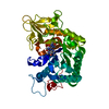
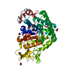
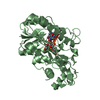
- PDB-6yn4: Structure of D169A/E171A double mutant of chitinase Chit42 from T... -
+ Open data

- Basic information
| Entry | Database: PDB / ID: 6yn4 | ||||||
|---|---|---|---|---|---|---|---|
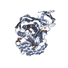
| Title | Structure of D169A/E171A double mutant of chitinase Chit42 from Trichoderma harzianum complexed with chitintetraose. | ||||||
 Components Components | Endochitinase 42 | ||||||
 Keywords Keywords | HYDROLASE / CHITINASE / HYDROLYTIC ENZYME / GLYCOSIL HYDROLASE / CHITIN / N-ACETYLGLUCOSAMINE / CHITINTETRAOSE / TETRAACETYL-CHITOTETRAOSE / COMPLEX / PREBIOTIC / OLIGOSACCHARIDE / CHITOOLOGOSACCHARIDE / TRICHODERMA HARZIANUM | ||||||
| Function / homology |  Function and homology information Function and homology informationendochitinase activity / chitinase / chitin catabolic process / chitin binding / polysaccharide catabolic process / extracellular region Similarity search - Function | ||||||
| Biological species |  Trichoderma harzianum (fungus) Trichoderma harzianum (fungus) | ||||||
| Method |  X-RAY DIFFRACTION / SYNCHROTRON / MOLECULAR REPLACEMENT / Resolution: 1.82 Å X-RAY DIFFRACTION / SYNCHROTRON / MOLECULAR REPLACEMENT / Resolution: 1.82 Å | ||||||
 Authors Authors | Jimenez-Ortega, E. / Sanz-Aparicio, J. | ||||||
 Citation Citation | Journal: Comput Struct Biotechnol J / Year: 2021 Title: Structural inspection and protein motions modelling of a fungal glycoside hydrolase family 18 chitinase by crystallography depicts a dynamic enzymatic mechanism Authors: Jimenez-Ortega, E. / Kidibule, P.E. / Fernandez-Lobato, M. / Sanz-Aparicio, J. | ||||||
| History |
|
- Structure visualization
| Structure viewer | Molecule: MolmilJmol/JSmol |
|---|
- Downloads & links
-Download
| PDBx/mmCIF format | 6yn4.cif.gz | 104.4 KB | Display | PDBx/mmCIF format |
|---|---|---|---|---|
| PDB format | pdb6yn4.ent.gz | 77.3 KB | Display | PDB format |
| PDBx/mmJSON format | 6yn4.json.gz | Tree view | PDBx/mmJSON format | |
| Others |  Other downloads Other downloads |
-Validation report
| Arichive directory | https://data.pdbj.org/pub/pdb/validation_reports/yn/6yn4ftp://data.pdbj.org/pub/pdb/validation_reports/yn/6yn4 | HTTPS FTP |
|---|
-Related structure data
| Related structure data |  6yljC  7akqC  6epbS S: Starting model for refinement C: citing same article ( |
|---|---|
| Similar structure data |


-Links
 PDBj
PDBj- Assembly
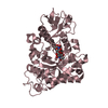
| Deposited unit | 
| ||||||||
|---|---|---|---|---|---|---|---|---|---|
| 1 |
| ||||||||
| Unit cell |
|
-Components
-Protein , 1 types, 1 molecules A
| #1: Protein | Mass: 45968.234 Da / Num. of mol.: 1 Source method: isolated from a genetically manipulated source Source: (gene. exp.) Trichoderma harzianum (fungus) / Gene: chit42 / Plasmid: pBluescript SK (+) / Production host: Komagataella phaffii GS115 (fungus) / References: UniProt: P48827, chitinase |
|---|
-Sugars , 2 types, 2 molecules
| #2: Polysaccharide | 2-acetamido-2-deoxy-beta-D-glucopyranose-(1-4)-2-acetamido-2-deoxy-beta-D-glucopyranose-(1-4)-2- ...2-acetamido-2-deoxy-beta-D-glucopyranose-(1-4)-2-acetamido-2-deoxy-beta-D-glucopyranose-(1-4)-2-acetamido-2-deoxy-beta-D-glucopyranose-(1-4)-2-acetamido-2-deoxy-beta-D-glucopyranose Source method: isolated from a genetically manipulated source |
|---|---|
| #3: Polysaccharide | 2-acetamido-2-deoxy-beta-D-glucopyranose-(1-4)-2-acetamido-2-deoxy-beta-D-glucopyranose Source method: isolated from a genetically manipulated source |
-Non-polymers , 4 types, 403 molecules 


| #4: Chemical | ChemComp-ACT /  Mass: 59.044 Da / Num. of mol.: 4 / Source method: obtained synthetically / Formula: C2H3O2 Mass: 59.044 Da / Num. of mol.: 4 / Source method: obtained synthetically / Formula: C2H3O2#5: Chemical |  Mass: 106.120 Da / Num. of mol.: 3 / Source method: obtained synthetically / Formula: C4H10O3 Mass: 106.120 Da / Num. of mol.: 3 / Source method: obtained synthetically / Formula: C4H10O3#6: Chemical | ChemComp-ZN /  Mass: 65.409 Da / Num. of mol.: 5 / Source method: obtained synthetically / Formula: Zn Mass: 65.409 Da / Num. of mol.: 5 / Source method: obtained synthetically / Formula: Zn#7: Water | ChemComp-HOH / | Mass: 18.015 Da / Num. of mol.: 391 / Source method: isolated from a natural source / Formula: H2O |
|---|
-Details
| Has ligand of interest | Y |
|---|
-Experimental details
-Experiment
| Experiment | Method: X-RAY DIFFRACTION / Number of used crystals: 1 |
|---|
- Sample preparation
| Crystal | Density Matthews: 2.47 Å3/Da / Density % sol: 50.27 % / Description: Bar |
|---|---|
| Crystal grow | Temperature: 291 K / Method: vapor diffusion, sitting drop / pH: 8 Details: 15 PEG 3000, 0.2 M Zinc Acetate, 0.1 M imidazole pH 8, 3% MPD Co-crystallization 2mM EDTA, 20mM chitintetraose. Cryoprotectant mother liquor supplemented with 20% PEG 400, 5 mM chitintetraose, 2 mM EDTA. |
-Data collection
| Diffraction | Mean temperature: 100 K / Serial crystal experiment: N |
|---|---|
| Diffraction source | Source: SYNCHROTRON / Site: ALBA  / Beamline: XALOC / Wavelength: 0.97924 Å / Beamline: XALOC / Wavelength: 0.97924 Å |
| Detector | Type: DECTRIS PILATUS 6M / Detector: PIXEL / Date: Sep 28, 2018 / Details: KB focusing mirrors |
| Radiation | Monochromator: Si(111) channel-cut, cryocooled / Protocol: SINGLE WAVELENGTH / Monochromatic (M) / Laue (L): M / Scattering type: x-ray |
| Radiation wavelength | Wavelength: 0.97924 Å / Relative weight: 1 |
| Reflection | Resolution: 1.82→48.38 Å / Num. obs: 38716 / % possible obs: 99.9 % / Redundancy: 6.5 % / CC1/2: 0.997 / Rmerge(I) obs: 0.107 / Rpim(I) all: 0.045 / Rrim(I) all: 0.117 / Net I/σ(I): 14.2 |
| Reflection shell | Resolution: 1.82→1.86 Å / Redundancy: 6.7 % / Rmerge(I) obs: 0.701 / Mean I/σ(I) obs: 4.8 / Num. unique obs: 2258 / CC1/2: 0.917 / Rpim(I) all: 0.286 / Rrim(I) all: 0.758 / % possible all: 100 |
- Processing
| Software |
| ||||||||||||||||||||||||||||||||||||||||||||||||||||||||||||
|---|---|---|---|---|---|---|---|---|---|---|---|---|---|---|---|---|---|---|---|---|---|---|---|---|---|---|---|---|---|---|---|---|---|---|---|---|---|---|---|---|---|---|---|---|---|---|---|---|---|---|---|---|---|---|---|---|---|---|---|---|---|
| Refinement | Method to determine structure: MOLECULAR REPLACEMENT Starting model: 6EPB Resolution: 1.82→48.38 Å / Cor.coef. Fo:Fc: 0.951 / Cor.coef. Fo:Fc free: 0.935 / SU B: 2.668 / SU ML: 0.082 / Cross valid method: THROUGHOUT / σ(F): 0 / ESU R: 0.133 / ESU R Free: 0.122 / Stereochemistry target values: MAXIMUM LIKELIHOOD Details: HYDROGENS HAVE BEEN ADDED IN THE RIDING POSITIONS U VALUES : REFINED INDIVIDUALLY
| ||||||||||||||||||||||||||||||||||||||||||||||||||||||||||||
| Solvent computation | Ion probe radii: 0.8 Å / Shrinkage radii: 0.8 Å / VDW probe radii: 1.2 Å / Solvent model: MASK | ||||||||||||||||||||||||||||||||||||||||||||||||||||||||||||
| Displacement parameters | Biso max: 52.82 Å2 / Biso mean: 15.511 Å2 / Biso min: 6.13 Å2
| ||||||||||||||||||||||||||||||||||||||||||||||||||||||||||||
| Refinement step | Cycle: final / Resolution: 1.82→48.38 Å
| ||||||||||||||||||||||||||||||||||||||||||||||||||||||||||||
| Refine LS restraints |
| ||||||||||||||||||||||||||||||||||||||||||||||||||||||||||||
| LS refinement shell | Resolution: 1.82→1.867 Å / Rfactor Rfree error: 0 / Total num. of bins used: 20
|
