 Movie
Movie Controller

[English] 日本語
 Yorodumi
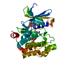
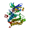
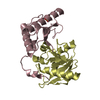
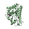
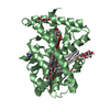
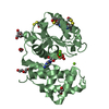
- PDB-6xz0: Crystal structure of spectinomycin adenyltransferase AAD(9) from ... -
+ Open data

- Basic information
| Entry | Database: PDB / ID: 6xz0 | |||||||||
|---|---|---|---|---|---|---|---|---|---|---|
| Title | Crystal structure of spectinomycin adenyltransferase AAD(9) from Enterococcus faecialis with spectinomycin | |||||||||
 Components Components | Streptomycin 3''-adenylyltransferase | |||||||||
 Keywords Keywords | TRANSFERASE / nucleotidyltransferase / antibiotic resistance | |||||||||
| Function / homology |  Function and homology information Function and homology information | |||||||||
| Biological species |   Enterococcus faecalis (bacteria) Enterococcus faecalis (bacteria) | |||||||||
| Method |  X-RAY DIFFRACTION / SYNCHROTRON / MOLECULAR REPLACEMENT / Resolution: 2.8 Å X-RAY DIFFRACTION / SYNCHROTRON / MOLECULAR REPLACEMENT / Resolution: 2.8 Å | |||||||||
 Authors Authors | Kanchugal P, S. / Selmer, M. | |||||||||
| Funding support |  Sweden, 2items Sweden, 2items
| |||||||||
 Citation Citation | Journal: Antimicrob.Agents Chemother. / Year: 2020 Title: Structural Recognition of Spectinomycin by Resistance Enzyme ANT(9) from Enterococcus faecalis. Authors: Kanchugal P, S. / Selmer, M. | |||||||||
| History |
|
- Structure visualization
| Structure viewer | Molecule: MolmilJmol/JSmol |
|---|
- Downloads & links
-Download
| PDBx/mmCIF format | 6xz0.cif.gz | 77.4 KB | Display | PDBx/mmCIF format |
|---|---|---|---|---|
| PDB format | pdb6xz0.ent.gz | 46.5 KB | Display | PDB format |
| PDBx/mmJSON format | 6xz0.json.gz | Tree view | PDBx/mmJSON format | |
| Others |  Other downloads Other downloads |
-Validation report
| Arichive directory | https://data.pdbj.org/pub/pdb/validation_reports/xz/6xz0ftp://data.pdbj.org/pub/pdb/validation_reports/xz/6xz0 | HTTPS FTP |
|---|
-Related structure data
| Related structure data | 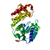 6sxjSC  6xxqC S: Starting model for refinement C: citing same article ( |
|---|---|
| Similar structure data |


-Links
 PDBj
PDBj


- Assembly
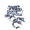
| Deposited unit | 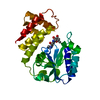
| ||||||||||||
|---|---|---|---|---|---|---|---|---|---|---|---|---|---|
| 1 |
| ||||||||||||
| Unit cell |
|
-Components
| #1: Protein | Mass: 30873.424 Da / Num. of mol.: 1 Source method: isolated from a genetically manipulated source Source: (gene. exp.) Enterococcus faecalis (bacteria) / Gene: spc, aad9 / Production host: References: UniProt: Q07448, streptomycin 3''-adenylyltransferase |
|---|---|
| #2: Chemical | ChemComp-SCM / 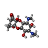  Mass: 332.350 Da / Num. of mol.: 1 / Source method: obtained synthetically / Formula: C14H24N2O7 / Feature type: SUBJECT OF INVESTIGATION Mass: 332.350 Da / Num. of mol.: 1 / Source method: obtained synthetically / Formula: C14H24N2O7 / Feature type: SUBJECT OF INVESTIGATION |
| Has ligand of interest | Y |
-Experimental details
-Experiment
| Experiment | Method: X-RAY DIFFRACTION / Number of used crystals: 1 |
|---|
- Sample preparation
| Crystal | Density Matthews: 2.46 Å3/Da / Density % sol: 60 % / Description: Hexagonal prism-shaped |
|---|---|
| Crystal grow | Temperature: 281 K / Method: vapor diffusion, sitting drop / pH: 8.5 Details: 10% w/v PEG 4000, 20% v/v glycerol, 0.02 M of each alcohol (0.2 M 1,6-hexanediol, 0.2 M 1-butanol, 0.2 M (RS)-1,2-propanediol, 0.2 M 2-propanol, 0.2 M 1,4-butanediol, 0.2 M 1,3-propanediol), ...Details: 10% w/v PEG 4000, 20% v/v glycerol, 0.02 M of each alcohol (0.2 M 1,6-hexanediol, 0.2 M 1-butanol, 0.2 M (RS)-1,2-propanediol, 0.2 M 2-propanol, 0.2 M 1,4-butanediol, 0.2 M 1,3-propanediol), 0.1 M bicine/Trizma base pH 8.5) were soaked with 10 mM ATP, 10 mM magnesium and spectinomycin powder for 30 seconds |
-Data collection
| Diffraction | Mean temperature: 100 K / Serial crystal experiment: N |
|---|---|
| Diffraction source | Source: SYNCHROTRON / Site: Diamond  / Beamline: I04 / Wavelength: 0.9795 Å / Beamline: I04 / Wavelength: 0.9795 Å |
| Detector | Type: DECTRIS EIGER2 XE 16M / Detector: PIXEL / Date: Apr 27, 2019 |
| Radiation | Protocol: SINGLE WAVELENGTH / Monochromatic (M) / Laue (L): M / Scattering type: x-ray |
| Radiation wavelength | Wavelength: 0.9795 Å / Relative weight: 1 |
| Reflection | Resolution: 2.8→49.8 Å / Num. obs: 7509 / % possible obs: 99.6 % / Redundancy: 22.9 % / Biso Wilson estimate: 69.18 Å2 / CC1/2: 0.9 / CC star: 1 / Rmerge(I) obs: 0.1 / Rrim(I) all: 0.1 / Net I/σ(I): 19.8 |
| Reflection shell | Resolution: 2.8→2.9 Å / Rmerge(I) obs: 1.5 / Num. unique obs: 751 / CC1/2: 0.9 / CC star: 0.9 / % possible all: 99.5 |
- Processing
| Software |
| ||||||||||||||||||||||||||||
|---|---|---|---|---|---|---|---|---|---|---|---|---|---|---|---|---|---|---|---|---|---|---|---|---|---|---|---|---|---|
| Refinement | Method to determine structure: MOLECULAR REPLACEMENT Starting model: 6SXJ Resolution: 2.8→49.78 Å / SU ML: 0.5886 / Cross valid method: FREE R-VALUE / σ(F): 1.35 / Phase error: 41.7612
| ||||||||||||||||||||||||||||
| Solvent computation | Shrinkage radii: 0.9 Å / VDW probe radii: 1.11 Å | ||||||||||||||||||||||||||||
| Displacement parameters | Biso mean: 75.88 Å2 | ||||||||||||||||||||||||||||
| Refinement step | Cycle: LAST / Resolution: 2.8→49.78 Å
| ||||||||||||||||||||||||||||
| Refine LS restraints |
| ||||||||||||||||||||||||||||
| LS refinement shell |
|
