 Movie
Movie Controller


+ Open data

- Basic information
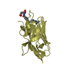
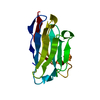
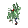
| Entry | Database: PDB / ID: 6xyf | ||||||
|---|---|---|---|---|---|---|---|
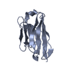
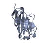
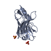
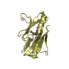
| Title | Nanobody 22 | ||||||
 Components Components | Nanobody 22 | ||||||
 Keywords Keywords | IMMUNE SYSTEM / Nanobody | ||||||
| Biological species |  | ||||||
| Method |  X-RAY DIFFRACTION / SYNCHROTRON / MOLECULAR REPLACEMENT / Resolution: 1.11097 Å X-RAY DIFFRACTION / SYNCHROTRON / MOLECULAR REPLACEMENT / Resolution: 1.11097 Å | ||||||
 Authors Authors | Pompidor, G. / Zimmermann, S. / Loew, C. / Schneider, T. | ||||||
 Citation Citation | Journal: To Be Published Title: Engineered nanobodies with a lanthanide binding motif for crystallographic phasing Authors: Pompidor, G. / Zimmermann, S. / Loew, C. / Schneider, T. | ||||||
| History |
|
- Structure visualization
| Structure viewer | Molecule:  MolmilJmol/JSmol MolmilJmol/JSmol |
|---|
- Downloads & links
-Download
| PDBx/mmCIF format | 6xyf.cif.gz | 96.2 KB | Display | PDBx/mmCIF format |
|---|---|---|---|---|
| PDB format | pdb6xyf.ent.gz | 70.6 KB | Display | PDB format |
| PDBx/mmJSON format | 6xyf.json.gz | Tree view | PDBx/mmJSON format | |
| Others |  Other downloads Other downloads |
-Validation report
| Arichive directory | https://data.pdbj.org/pub/pdb/validation_reports/xy/6xyfftp://data.pdbj.org/pub/pdb/validation_reports/xy/6xyf | HTTPS FTP |
|---|
-Related structure data
| Related structure data |  6xymC  6xzfC  6y0eC  6y1rC  4i13S S: Starting model for refinement C: citing same article ( |
|---|---|
| Similar structure data |


-Links
 PDBj
PDBj


- Assembly
| Deposited unit | 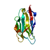
| ||||||||||
|---|---|---|---|---|---|---|---|---|---|---|---|
| 1 |
| ||||||||||
| Unit cell |
|
-Components
| #1: Antibody | Mass: 14276.796 Da / Num. of mol.: 1 Source method: isolated from a genetically manipulated source Source: (gene. exp.) Production host:  |
|---|---|
| #2: Chemical | ChemComp-NA /   Mass: 22.990 Da / Num. of mol.: 1 / Source method: obtained synthetically / Formula: Na Mass: 22.990 Da / Num. of mol.: 1 / Source method: obtained synthetically / Formula: Na |
| #3: Water | ChemComp-HOH /  Mass: 18.015 Da / Num. of mol.: 143 / Source method: isolated from a natural source / Formula: H2O Mass: 18.015 Da / Num. of mol.: 143 / Source method: isolated from a natural source / Formula: H2O |
| Has ligand of interest | N |
| Has protein modification | Y |
-Experimental details
-Experiment
| Experiment | Method: X-RAY DIFFRACTION / Number of used crystals: 1 |
|---|
- Sample preparation
| Crystal | Density Matthews: 1.82 Å3/Da / Density % sol: 32.57 % |
|---|---|
| Crystal grow | Temperature: 298 K / Method: vapor diffusion, hanging drop Details: 0.1 M Bis Tris Propane 100 mM Na/K Phosphate 30 % PEG 3350 |
-Data collection
| Diffraction | Mean temperature: 100 K / Serial crystal experiment: N | ||||||||||||||||||||||||
|---|---|---|---|---|---|---|---|---|---|---|---|---|---|---|---|---|---|---|---|---|---|---|---|---|---|
| Diffraction source | Source: SYNCHROTRON / Site: PETRA III, EMBL c/o DESY  / Beamline: P14 (MX2) / Wavelength: 0.9484 Å / Beamline: P14 (MX2) / Wavelength: 0.9484 Å | ||||||||||||||||||||||||
| Detector | Type: DECTRIS PILATUS 6M / Detector: PIXEL / Date: May 30, 2015 | ||||||||||||||||||||||||
| Radiation | Protocol: SINGLE WAVELENGTH / Monochromatic (M) / Laue (L): M / Scattering type: x-ray | ||||||||||||||||||||||||
| Radiation wavelength | Wavelength: 0.9484 Å / Relative weight: 1 | ||||||||||||||||||||||||
| Reflection | Resolution: 1.11→30.01 Å / Num. obs: 39057 / % possible obs: 96.3 % / Redundancy: 12.7 % / CC1/2: 0.997 / Rmerge(I) obs: 0.13 / Rpim(I) all: 0.037 / Rrim(I) all: 0.135 / Net I/σ(I): 10.2 / Num. measured all: 495655 / Scaling rejects: 668 | ||||||||||||||||||||||||
| Reflection shell | Diffraction-ID: 1
|
- Processing
| Software |
| ||||||||||||||||||||||||
|---|---|---|---|---|---|---|---|---|---|---|---|---|---|---|---|---|---|---|---|---|---|---|---|---|---|
| Refinement | Method to determine structure: MOLECULAR REPLACEMENT Starting model: 4i13.pdb Resolution: 1.11097→30.0062 Å / Cross valid method: FREE R-VALUE
| ||||||||||||||||||||||||
| Displacement parameters | Biso mean: 13.7 Å2 | ||||||||||||||||||||||||
| Refinement step | Cycle: LAST / Resolution: 1.11097→30.0062 Å
| ||||||||||||||||||||||||
| Refine LS restraints |
|
