 Movie
Movie Controller

[English] 日本語
 Yorodumi
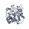
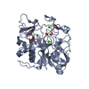
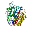
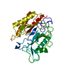
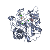
- PDB-6xtp: Crystal structure reveals non-coordinative binding of O2 to the c... -
+ Open data

- Basic information
| Entry | Database: PDB / ID: 6xtp | |||||||||
|---|---|---|---|---|---|---|---|---|---|---|
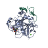
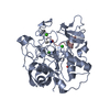
| Title | Crystal structure reveals non-coordinative binding of O2 to the copper center of the formylglycine-generating enzyme - FGE:Cu:S:O2-1a complex | |||||||||
 Components Components |
| |||||||||
 Keywords Keywords | TRANSFERASE / Formylglycine-generating enzyme / complex / substrate analog / copper | |||||||||
| Function / homology |  Function and homology information Function and homology informationformylglycine-generating enzyme / formylglycine-generating oxidase activity / protein oxidation / cuprous ion binding / post-translational protein modification / metal ion binding Similarity search - Function | |||||||||
| Biological species |  Thermomonospora curvata (bacteria)Thermomonospora curvata DSM 43183 (bacteria) Thermomonospora curvata (bacteria)Thermomonospora curvata DSM 43183 (bacteria) | |||||||||
| Method |  X-RAY DIFFRACTION / SYNCHROTRON / MOLECULAR REPLACEMENT / Resolution: 1.8 Å X-RAY DIFFRACTION / SYNCHROTRON / MOLECULAR REPLACEMENT / Resolution: 1.8 Å | |||||||||
 Authors Authors | Leisinger, F. / Seebeck, F.P. | |||||||||
| Funding support |  Switzerland, European Union, 2items Switzerland, European Union, 2items
| |||||||||
 Citation Citation | Journal: Angew.Chem.Int.Ed.Engl. / Year: 2020 Title: Non-Coordinative Binding of O2 at the Active Center of a Copper-Dependent Enzyme Authors: Leisinger, F. / Miarzlou, D.A. / Seebeck, F.P. | |||||||||
| History |
|
- Structure visualization
| Structure viewer | Molecule: MolmilJmol/JSmol |
|---|
- Downloads & links
-Download
| PDBx/mmCIF format | 6xtp.cif.gz | 81 KB | Display | PDBx/mmCIF format |
|---|---|---|---|---|
| PDB format | pdb6xtp.ent.gz | 57.3 KB | Display | PDB format |
| PDBx/mmJSON format | 6xtp.json.gz | Tree view | PDBx/mmJSON format | |
| Others |  Other downloads Other downloads |
-Validation report
| Arichive directory | https://data.pdbj.org/pub/pdb/validation_reports/xt/6xtpftp://data.pdbj.org/pub/pdb/validation_reports/xt/6xtp | HTTPS FTP |
|---|
-Related structure data
| Related structure data | 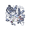 6xtlC 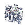 6xtmC  6xtnC  6xtoC  6xtqC 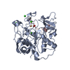 6xtrC  6xtsC  6s07S S: Starting model for refinement C: citing same article ( |
|---|---|
| Similar structure data |


-Links
 PDBj
PDBj


- Assembly
| Deposited unit | 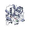
| ||||||||
|---|---|---|---|---|---|---|---|---|---|
| 1 |
| ||||||||
| Unit cell |
|
-Components
-Protein / Protein/peptide , 2 types, 2 molecules AC
| #1: Protein | Mass: 33336.852 Da / Num. of mol.: 1 Source method: isolated from a genetically manipulated source Source: (gene. exp.) Thermomonospora curvata (strain ATCC 19995 / DSM 43183 / JCM 3096 / NBRC 15933 / NCIMB 10081 / Henssen B9) (bacteria)Gene: Tcur_4811 / Plasmid: pET19 / Production host: References: UniProt: D1A7C3, formylglycine-generating enzyme |
|---|---|
| #2: Protein/peptide | Mass: 1387.625 Da / Num. of mol.: 1 / Source method: obtained synthetically / Details: modified sulfatase sequence motif Source: (synth.) Thermomonospora curvata DSM 43183 (bacteria)References: UniProt: D1ADF2*PLUS |
-Non-polymers , 5 types, 199 molecules 


| #3: Chemical | ChemComp-CU1 /  Mass: 63.546 Da / Num. of mol.: 1 / Source method: obtained synthetically / Formula: Cu Mass: 63.546 Da / Num. of mol.: 1 / Source method: obtained synthetically / Formula: Cu | ||||||
|---|---|---|---|---|---|---|---|
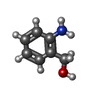
| #4: Chemical |  Mass: 40.078 Da / Num. of mol.: 2 / Source method: obtained synthetically / Formula: Ca Mass: 40.078 Da / Num. of mol.: 2 / Source method: obtained synthetically / Formula: Ca#5: Chemical | ChemComp-OXY / |  Mass: 31.999 Da / Num. of mol.: 1 / Source method: obtained synthetically / Formula: O2 Mass: 31.999 Da / Num. of mol.: 1 / Source method: obtained synthetically / Formula: O2#6: Chemical | ChemComp-SOA / |  Mass: 123.152 Da / Num. of mol.: 1 / Source method: obtained synthetically / Formula: C7H9NO Mass: 123.152 Da / Num. of mol.: 1 / Source method: obtained synthetically / Formula: C7H9NO#7: Water | ChemComp-HOH / | Mass: 18.015 Da / Num. of mol.: 194 / Source method: isolated from a natural source / Formula: H2O |
-Experimental details
-Experiment
| Experiment | Method: X-RAY DIFFRACTION / Number of used crystals: 1 |
|---|
- Sample preparation
| Crystal | Density Matthews: 2.22 Å3/Da / Density % sol: 44.49 % |
|---|---|
| Crystal grow | Temperature: 293 K / Method: vapor diffusion, sitting drop / pH: 7 / Details: 7-12 % PEG 8000, 0.2-0.3 M MgCl2, Tris-HCl |
-Data collection
| Diffraction | Mean temperature: 100 K / Serial crystal experiment: N | ||||||||||||||||||||||||||||||
|---|---|---|---|---|---|---|---|---|---|---|---|---|---|---|---|---|---|---|---|---|---|---|---|---|---|---|---|---|---|---|---|
| Diffraction source | Source: SYNCHROTRON / Site: SLS / Beamline: X06SA / Wavelength: 1.0006777109999 Å | ||||||||||||||||||||||||||||||
| Detector | Type: DECTRIS PILATUS 2M-F / Detector: PIXEL / Date: Oct 5, 2019 | ||||||||||||||||||||||||||||||
| Radiation | Protocol: SINGLE WAVELENGTH / Monochromatic (M) / Laue (L): M / Scattering type: x-ray | ||||||||||||||||||||||||||||||
| Radiation wavelength | Wavelength: 1.0006777109999 Å / Relative weight: 1 | ||||||||||||||||||||||||||||||
| Reflection | Resolution: 1.8→45.69 Å / Num. obs: 26230 / % possible obs: 89.5 % / Redundancy: 10.4 % / Biso Wilson estimate: 13.64 Å2 / CC1/2: 0.997 / Rmerge(I) obs: 0.092 / Rpim(I) all: 0.031 / Rrim(I) all: 0.098 / Net I/σ(I): 16.3 / Num. measured all: 272094 / Scaling rejects: 67 | ||||||||||||||||||||||||||||||
| Reflection shell | Diffraction-ID: 1
|
- Processing
| Software |
| ||||||||||||||||||||||||||||||||||||||||||||||||||||||
|---|---|---|---|---|---|---|---|---|---|---|---|---|---|---|---|---|---|---|---|---|---|---|---|---|---|---|---|---|---|---|---|---|---|---|---|---|---|---|---|---|---|---|---|---|---|---|---|---|---|---|---|---|---|---|---|
| Refinement | Method to determine structure: MOLECULAR REPLACEMENT Starting model: 6S07 Resolution: 1.8→45.688 Å / SU ML: 0.3 / Cross valid method: THROUGHOUT / σ(F): 1.33 / Phase error: 33.64
| ||||||||||||||||||||||||||||||||||||||||||||||||||||||
| Solvent computation | Shrinkage radii: 0.9 Å / VDW probe radii: 1.11 Å | ||||||||||||||||||||||||||||||||||||||||||||||||||||||
| Displacement parameters | Biso max: 50.92 Å2 / Biso mean: 21.7826 Å2 / Biso min: 8.57 Å2 | ||||||||||||||||||||||||||||||||||||||||||||||||||||||
| Refinement step | Cycle: final / Resolution: 1.8→45.688 Å
| ||||||||||||||||||||||||||||||||||||||||||||||||||||||
| Refine LS restraints |
| ||||||||||||||||||||||||||||||||||||||||||||||||||||||
| LS refinement shell | Refine-ID: X-RAY DIFFRACTION / Rfactor Rfree error: 0
|
