 Movie
Movie Controller

[English] 日本語
 Yorodumi
- PDB-6xt1: The structure of the M60 catalytic domain from Clostridium perfri... -
+ Open data

- Basic information
| Entry | Database: PDB / ID: 6xt1 | |||||||||
|---|---|---|---|---|---|---|---|---|---|---|
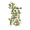
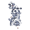
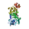
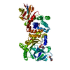
| Title | The structure of the M60 catalytic domain from Clostridium perfringens ZmpC in complex the sialyl T antigen | |||||||||
 Components Components | ZmpC Glycopeptidase | |||||||||
 Keywords Keywords | HYDROLASE / glycopeptidase | |||||||||
| Function / homology |  Function and homology information Function and homology information | |||||||||
| Biological species |   Clostridium perfringens (bacteria) Clostridium perfringens (bacteria) | |||||||||
| Method |  X-RAY DIFFRACTION / MOLECULAR REPLACEMENT / molecular replacement / Resolution: 2.49 Å X-RAY DIFFRACTION / MOLECULAR REPLACEMENT / molecular replacement / Resolution: 2.49 Å | |||||||||
 Authors Authors | Pluvinage, B. / Boraston, A.B. | |||||||||
| Funding support |  Canada, 2items Canada, 2items
| |||||||||
 Citation Citation | Journal: Proc.Natl.Acad.Sci.USA / Year: 2021 Title: Molecular insights into architecturally complex glycopeptidases Authors: Pluvinage, B. / Ficko-Blean, E. / Noach, I. / Stuart, C. / Thompson, N. / McClure, H. / Buenbrazo, N. / Wakarchuck, W. / Boraston, A.B. | |||||||||
| History |
|
- Structure visualization
| Structure viewer | Molecule: MolmilJmol/JSmol |
|---|
- Downloads & links
-Download
| PDBx/mmCIF format | 6xt1.cif.gz | 416.3 KB | Display | PDBx/mmCIF format |
|---|---|---|---|---|
| PDB format | pdb6xt1.ent.gz | 336 KB | Display | PDB format |
| PDBx/mmJSON format | 6xt1.json.gz | Tree view | PDBx/mmJSON format | |
| Others |  Other downloads Other downloads |
-Validation report
| Arichive directory | https://data.pdbj.org/pub/pdb/validation_reports/xt/6xt1ftp://data.pdbj.org/pub/pdb/validation_reports/xt/6xt1 | HTTPS FTP |
|---|
-Related structure data
| Related structure data |  6xsxC  6xszSC S: Starting model for refinement C: citing same article ( |
|---|---|
| Similar structure data |


-Links
 PDBj
PDBj

- Assembly
| Deposited unit | 
| ||||||||
|---|---|---|---|---|---|---|---|---|---|
| 1 | 
| ||||||||
| 2 | 
| ||||||||
| 3 | 
| ||||||||
| 4 | 
| ||||||||
| Unit cell |
|
-Components
-Protein / Sugars , 2 types, 6 molecules ABCD
| #1: Protein | Mass: 60238.398 Da / Num. of mol.: 4 / Fragment: M60 catalytic domain Source method: isolated from a genetically manipulated source Source: (gene. exp.) Clostridium perfringens (bacteria) / Gene: CP4_3468 / Plasmid: pET28a / Production host: #2: Polysaccharide | |
|---|
-Non-polymers , 4 types, 537 molecules 


| #3: Chemical | ChemComp-ZN /  Mass: 65.409 Da / Num. of mol.: 4 / Source method: obtained synthetically / Formula: Zn Mass: 65.409 Da / Num. of mol.: 4 / Source method: obtained synthetically / Formula: Zn#4: Chemical | ChemComp-EDO /  Mass: 62.068 Da / Num. of mol.: 16 / Source method: obtained synthetically / Formula: C2H6O2 Mass: 62.068 Da / Num. of mol.: 16 / Source method: obtained synthetically / Formula: C2H6O2#5: Chemical |  Type: L-peptide linking / Mass: 105.093 Da / Num. of mol.: 2 / Source method: obtained synthetically / Formula: C3H7NO3 Type: L-peptide linking / Mass: 105.093 Da / Num. of mol.: 2 / Source method: obtained synthetically / Formula: C3H7NO3#6: Water | ChemComp-HOH / | Mass: 18.015 Da / Num. of mol.: 515 / Source method: isolated from a natural source / Formula: H2O |
|---|
-Details
| Has ligand of interest | Y |
|---|
-Experimental details
-Experiment
| Experiment | Method: X-RAY DIFFRACTION / Number of used crystals: 1 |
|---|
- Sample preparation
| Crystal | Density Matthews: 2.72 Å3/Da / Density % sol: 54.75 % |
|---|---|
| Crystal grow | Temperature: 291 K / Method: vapor diffusion, hanging drop / pH: 5.8 / Details: 7.6% Tascimate, 19% PEG 3350 |
-Data collection
| Diffraction | Mean temperature: 100 K / Serial crystal experiment: N |
|---|---|
| Diffraction source | Source: ROTATING ANODE / Type: RIGAKU MICROMAX-002 / Wavelength: 1.54187 Å |
| Detector | Type: DECTRIS PILATUS 200K / Detector: PIXEL / Date: Sep 7, 2016 |
| Radiation | Protocol: SINGLE WAVELENGTH / Monochromatic (M) / Laue (L): M / Scattering type: x-ray |
| Radiation wavelength | Wavelength: 1.54187 Å / Relative weight: 1 |
| Reflection | Resolution: 2.49→40 Å / Num. obs: 89691 / % possible obs: 99.9 % / Redundancy: 4.2 % / CC1/2: 0.988 / Rmerge(I) obs: 0.109 / Rpim(I) all: 0.051 / Net I/σ(I): 15.3 |
| Reflection shell | Resolution: 2.5→2.54 Å / Redundancy: 4 % / Rmerge(I) obs: 0.589 / Mean I/σ(I) obs: 2.3 / Num. unique obs: 4505 / CC1/2: 0.899 / Rpim(I) all: 0.317 / % possible all: 100 |
-Phasing
| Phasing | Method: molecular replacement |
|---|
- Processing
| Software |
| |||||||||||||||||||||||||||||||||||||||||||||
|---|---|---|---|---|---|---|---|---|---|---|---|---|---|---|---|---|---|---|---|---|---|---|---|---|---|---|---|---|---|---|---|---|---|---|---|---|---|---|---|---|---|---|---|---|---|---|
| Refinement | Method to determine structure: MOLECULAR REPLACEMENT Starting model: 6XSZ Resolution: 2.49→39.07 Å / Cor.coef. Fo:Fc: 0.93 / Cor.coef. Fo:Fc free: 0.893 / SU B: 11.328 / SU ML: 0.241 / Cross valid method: THROUGHOUT / σ(F): 0 / ESU R: 0.628 / ESU R Free: 0.324 / Stereochemistry target values: MAXIMUM LIKELIHOOD / Details: U VALUES : REFINED INDIVIDUALLY
| |||||||||||||||||||||||||||||||||||||||||||||
| Solvent computation | Ion probe radii: 0.8 Å / Shrinkage radii: 0.8 Å / VDW probe radii: 1.2 Å / Solvent model: MASK | |||||||||||||||||||||||||||||||||||||||||||||
| Displacement parameters | Biso max: 100.49 Å2 / Biso mean: 41.615 Å2 / Biso min: 20.23 Å2
| |||||||||||||||||||||||||||||||||||||||||||||
| Refinement step | Cycle: final / Resolution: 2.49→39.07 Å
| |||||||||||||||||||||||||||||||||||||||||||||
| Refine LS restraints |
| |||||||||||||||||||||||||||||||||||||||||||||
| LS refinement shell | Resolution: 2.494→2.559 Å / Rfactor Rfree error: 0 / Total num. of bins used: 20
|
