 Movie
Movie Controller

[English] 日本語
 Yorodumi
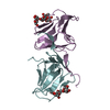
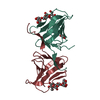
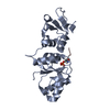
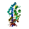
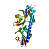
- PDB-6xp8: The crystal structure of TfuA involved in peptide backbone thioam... -
+ Open data

- Basic information
| Entry | Database: PDB / ID: 6xp8 | ||||||
|---|---|---|---|---|---|---|---|
| Title | The crystal structure of TfuA involved in peptide backbone thioamidation from Methanosarcina acetivorans | ||||||
 Components Components | TfuA domain-containing protein | ||||||
 Keywords Keywords | BIOSYNTHETIC PROTEIN / YcaO / TfuA / thioamidation | ||||||
| Function / homology | TfuA-like, core / TfuA-like protein / TfuA-like core domain-containing protein Function and homology information Function and homology information | ||||||
| Biological species |  Methanosarcina acetivorans (archaea) Methanosarcina acetivorans (archaea) | ||||||
| Method |  X-RAY DIFFRACTION / SYNCHROTRON / SAD / Resolution: 1.65 Å X-RAY DIFFRACTION / SYNCHROTRON / SAD / Resolution: 1.65 Å | ||||||
 Authors Authors | Dong, S.-H. / Nair, S.K. | ||||||
 Citation Citation | Journal: Nat.Chem.Biol. / Year: 2021 Title: Functional elucidation of TfuA in peptide backbone thioamidation. Authors: Liu, A. / Si, Y. / Dong, S.H. / Mahanta, N. / Penkala, H.N. / Nair, S.K. / Mitchell, D.A. | ||||||
| History |
|
- Structure visualization
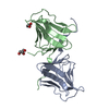
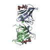
| Structure viewer | Molecule: MolmilJmol/JSmol |
|---|
- Downloads & links
-Download
| PDBx/mmCIF format | 6xp8.cif.gz | 60.4 KB | Display | PDBx/mmCIF format |
|---|---|---|---|---|
| PDB format | pdb6xp8.ent.gz | 42.3 KB | Display | PDB format |
| PDBx/mmJSON format | 6xp8.json.gz | Tree view | PDBx/mmJSON format | |
| Others |  Other downloads Other downloads |
-Validation report
| Arichive directory | https://data.pdbj.org/pub/pdb/validation_reports/xp/6xp8ftp://data.pdbj.org/pub/pdb/validation_reports/xp/6xp8 | HTTPS FTP |
|---|
-Related structure data
| Similar structure data |
|---|


-Links
 PDBj
PDBj- Assembly
| Deposited unit | 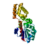
| |||||||||
|---|---|---|---|---|---|---|---|---|---|---|
| 1 |
| |||||||||
| Unit cell |
| |||||||||
| Components on special symmetry positions |
|
-Components
| #1: Protein | Mass: 23887.324 Da / Num. of mol.: 1 Source method: isolated from a genetically manipulated source Source: (gene. exp.) Methanosarcina acetivorans (archaea) / Production host:  |
|---|---|
| #2: Water | ChemComp-HOH /  Mass: 18.015 Da / Num. of mol.: 238 / Source method: isolated from a natural source / Formula: H2O Mass: 18.015 Da / Num. of mol.: 238 / Source method: isolated from a natural source / Formula: H2O |
-Experimental details
-Experiment
| Experiment | Method: X-RAY DIFFRACTION / Number of used crystals: 1 |
|---|
- Sample preparation
| Crystal | Density Matthews: 2.03 Å3/Da / Density % sol: 39.51 % |
|---|---|
| Crystal grow | Temperature: 282 K / Method: vapor diffusion, hanging drop Details: 100 mM sodium citrate tribasic/citric acid pH 5.5 and 40% (v/v) PEG 600 |
-Data collection
| Diffraction | Mean temperature: 80 K / Serial crystal experiment: N |
|---|---|
| Diffraction source | Source: SYNCHROTRON / Site: APS  / Beamline: 21-ID-F / Wavelength: 0.97872 Å / Beamline: 21-ID-F / Wavelength: 0.97872 Å |
| Detector | Type: RAYONIX MX300HE / Detector: CCD / Date: Jul 18, 2018 |
| Radiation | Protocol: SINGLE WAVELENGTH / Monochromatic (M) / Laue (L): M / Scattering type: x-ray |
| Radiation wavelength | Wavelength: 0.97872 Å / Relative weight: 1 |
| Reflection | Resolution: 1.65→25 Å / Num. obs: 23292 / % possible obs: 99.8 % / Redundancy: 7.4 % / CC1/2: 0.999 / Net I/σ(I): 21.4 |
| Reflection shell | Resolution: 1.65→10 Å / Num. unique obs: 22138 / CC1/2: 0.787 |
- Processing
| Software |
| |||||||||||||||||||||||||||||||||||||||||||||
|---|---|---|---|---|---|---|---|---|---|---|---|---|---|---|---|---|---|---|---|---|---|---|---|---|---|---|---|---|---|---|---|---|---|---|---|---|---|---|---|---|---|---|---|---|---|---|
| Refinement | Method to determine structure: SAD / Resolution: 1.65→25 Å / Cor.coef. Fo:Fc: 0.959 / Cor.coef. Fo:Fc free: 0.934 / SU B: 2.557 / SU ML: 0.086 / Cross valid method: THROUGHOUT / σ(F): 0 / ESU R: 0.12 / ESU R Free: 0.116 / Stereochemistry target values: MAXIMUM LIKELIHOOD Details: HYDROGENS HAVE BEEN USED IF PRESENT IN THE INPUT U VALUES : REFINED INDIVIDUALLY
| |||||||||||||||||||||||||||||||||||||||||||||
| Solvent computation | Ion probe radii: 0.8 Å / Shrinkage radii: 0.8 Å / VDW probe radii: 1.2 Å / Solvent model: MASK | |||||||||||||||||||||||||||||||||||||||||||||
| Displacement parameters | Biso max: 50.65 Å2 / Biso mean: 22.01 Å2 / Biso min: 12.33 Å2
| |||||||||||||||||||||||||||||||||||||||||||||
| Refinement step | Cycle: final / Resolution: 1.65→25 Å
| |||||||||||||||||||||||||||||||||||||||||||||
| Refine LS restraints |
| |||||||||||||||||||||||||||||||||||||||||||||
| LS refinement shell | Resolution: 1.652→1.695 Å / Rfactor Rfree error: 0
|
