 Movie
Movie Controller


+ Open data

- Basic information
| Entry | Database: PDB / ID: 6wpa | ||||||
|---|---|---|---|---|---|---|---|
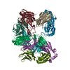
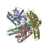
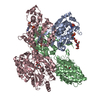
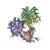
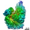
| Title | Structure of AvaR1 bound to DNA half-site | ||||||
 Components Components |
| ||||||
 Keywords Keywords | DNA BINDING PROTEIN/DNA / Transcriptional Repressor / DNA oligonucleotide / DNA-Protein biniding / DNA BINDING PROTEIN / DNA BINDING PROTEIN-DNA complex | ||||||
| Function / homology |  Function and homology information Function and homology information | ||||||
| Biological species |  Streptomyces avermitilis (bacteria)Streptomyces avermitilis MA-4680 = NBRC 14893 (bacteria) Streptomyces avermitilis (bacteria)Streptomyces avermitilis MA-4680 = NBRC 14893 (bacteria) | ||||||
| Method |  X-RAY DIFFRACTION / SYNCHROTRON / MOLECULAR REPLACEMENT / Resolution: 3.09 Å X-RAY DIFFRACTION / SYNCHROTRON / MOLECULAR REPLACEMENT / Resolution: 3.09 Å | ||||||
 Authors Authors | Kapoor, I. / Olivares, P.J. / Nair, S.K. | ||||||
| Funding support |  United States, 1items United States, 1items
| ||||||
 Citation Citation | Journal: Elife / Year: 2020 Title: Biochemical basis for the regulation of biosynthesis of antiparasitics by bacterial hormones. Authors: Kapoor, I. / Olivares, P. / Nair, S.K. | ||||||
| History |
|
- Structure visualization
| Structure viewer | Molecule: MolmilJmol/JSmol |
|---|
- Downloads & links
-Download
| PDBx/mmCIF format | 6wpa.cif.gz | 366.1 KB | Display | PDBx/mmCIF format |
|---|---|---|---|---|
| PDB format | pdb6wpa.ent.gz | 292.7 KB | Display | PDB format |
| PDBx/mmJSON format | 6wpa.json.gz | Tree view | PDBx/mmJSON format | |
| Others |  Other downloads Other downloads |
-Validation report
| Arichive directory | https://data.pdbj.org/pub/pdb/validation_reports/wp/6wpaftp://data.pdbj.org/pub/pdb/validation_reports/wp/6wpa | HTTPS FTP |
|---|
-Related structure data
| Related structure data |  6wp7SC 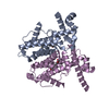 6wp9C S: Starting model for refinement C: citing same article ( |
|---|---|
| Similar structure data |


-Links
 PDBj
PDBj


- Assembly
| Deposited unit | 
| ||||||||
|---|---|---|---|---|---|---|---|---|---|
| 1 | 
| ||||||||
| 2 | 
| ||||||||
| Unit cell |
|
-Components
| #1: Protein | Mass: 27037.654 Da / Num. of mol.: 8 Source method: isolated from a genetically manipulated source Details: AvaR1 bound to DNA oligonucleotide through its DNA binding domain Source: (gene. exp.) Streptomyces avermitilis (bacteria)Strain: ATCC 31267 / DSM 46492 / JCM 5070 / NBRC 14893 / NCIMB 12804 / NRRL 8165 / MA-4680 Gene: avaR1, SAVERM_3705 / Plasmid: Rosetta Production host: References: UniProt: Q82H41 #2: DNA chain | Mass: 8604.565 Da / Num. of mol.: 2 / Source method: obtained synthetically / Details: Palindromic double stranded DNA (28-MER) Source: (synth.) Streptomyces avermitilis MA-4680 = NBRC 14893 (bacteria) |
|---|
-Experimental details
-Experiment
| Experiment | Method: X-RAY DIFFRACTION / Number of used crystals: 1 |
|---|
- Sample preparation
| Crystal | Density Matthews: 3.43 Å3/Da / Density % sol: 64.17 % |
|---|---|
| Crystal grow | Temperature: 277 K / Method: vapor diffusion, hanging drop / pH: 6.5 Details: 0.2 M Potassium chloride, 0.01 M Magnesium chloride hexahydrate, 0.05 M Sodium cacodylate trihydrate pH 6.5, 10% w/v Polyethylene glycol 4000 with additive 40 % Formamide |
-Data collection
| Diffraction | Mean temperature: 100 K / Serial crystal experiment: N |
|---|---|
| Diffraction source | Source: SYNCHROTRON / Site: APS / Beamline: 21-ID-D / Wavelength: 0.97872 Å |
| Detector | Type: MARMOSAIC 300 mm CCD / Detector: CCD / Date: Apr 14, 2018 |
| Radiation | Protocol: SINGLE WAVELENGTH / Monochromatic (M) / Laue (L): M / Scattering type: x-ray |
| Radiation wavelength | Wavelength: 0.97872 Å / Relative weight: 1 |
| Reflection | Resolution: 3.08→25 Å / Num. obs: 55544 / % possible obs: 100 % / Redundancy: 15.1 % / CC1/2: 0.999 / Rmerge(I) obs: 0.077 / Rpim(I) all: 0.021 / Rrim(I) all: 0.079 / Net I/σ(I): 25.9 |
| Reflection shell | Resolution: 3.09→3.2 Å / Num. unique obs: 5510 / CC1/2: 0.999 / Rpim(I) all: 0.338 |
- Processing
| Software |
| |||||||||||||||||||||||||||||||||||||||||||||
|---|---|---|---|---|---|---|---|---|---|---|---|---|---|---|---|---|---|---|---|---|---|---|---|---|---|---|---|---|---|---|---|---|---|---|---|---|---|---|---|---|---|---|---|---|---|---|
| Refinement | Method to determine structure: MOLECULAR REPLACEMENT Starting model: 6WP7 Resolution: 3.09→25 Å / Cor.coef. Fo:Fc: 0.949 / Cor.coef. Fo:Fc free: 0.923 / SU B: 20.645 / SU ML: 0.352 / Cross valid method: THROUGHOUT / σ(F): 0 / ESU R Free: 0.434 / Stereochemistry target values: MAXIMUM LIKELIHOOD Details: HYDROGENS HAVE BEEN USED IF PRESENT IN THE INPUT U VALUES : REFINED INDIVIDUALLY
| |||||||||||||||||||||||||||||||||||||||||||||
| Solvent computation | Ion probe radii: 0.8 Å / Shrinkage radii: 0.8 Å / VDW probe radii: 1.2 Å / Solvent model: MASK | |||||||||||||||||||||||||||||||||||||||||||||
| Displacement parameters | Biso max: 213.84 Å2 / Biso mean: 106.635 Å2 / Biso min: 48.06 Å2
| |||||||||||||||||||||||||||||||||||||||||||||
| Refinement step | Cycle: final / Resolution: 3.09→25 Å
| |||||||||||||||||||||||||||||||||||||||||||||
| Refine LS restraints |
| |||||||||||||||||||||||||||||||||||||||||||||
| LS refinement shell | Resolution: 3.09→3.165 Å / Rfactor Rfree error: 0
|
