 Movie
Movie Controller

[English] 日本語
 Yorodumi
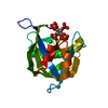
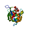
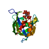
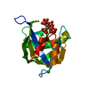
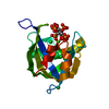
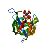
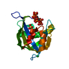
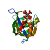
- PDB-6woa: Diphosphoinositol polyphosphate phosphohydrolase 1 (DIPP1/NUDT3) ... -
+ Open data

- Basic information
| Entry | Database: PDB / ID: 6woa | ||||||
|---|---|---|---|---|---|---|---|
| Title | Diphosphoinositol polyphosphate phosphohydrolase 1 (DIPP1/NUDT3) in complex with 2-Diphosphoinositol pentakisphosphate (2-IP7), Mg, and Fluoride ion | ||||||
 Components Components | Diphosphoinositol polyphosphate phosphohydrolase 1 | ||||||
 Keywords Keywords | HYDROLASE / phosphatase / nudix / catalysis mechanism / Substrate Specificity / inositol / inositol pyrophosphate | ||||||
| Function / homology |  Function and homology information Function and homology informationinositol diphosphate pentakisphosphate diphosphatase activity / diphosphoinositol polyphosphate catabolic process / inositol diphosphate tetrakisphosphate diphosphatase activity / endopolyphosphatase / diadenosine polyphosphate catabolic process / 5'-(N7-methyl 5'-triphosphoguanosine)-[mRNA] diphosphatase / bis(5'-adenosyl)-hexaphosphatase activity / inositol-3,5-bisdiphosphate-2,3,4,6-tetrakisphosphate 5-diphosphatase activity / diadenosine pentaphosphate catabolic process / diadenosine hexaphosphate catabolic process ...inositol diphosphate pentakisphosphate diphosphatase activity / diphosphoinositol polyphosphate catabolic process / inositol diphosphate tetrakisphosphate diphosphatase activity / endopolyphosphatase / diadenosine polyphosphate catabolic process / 5'-(N7-methyl 5'-triphosphoguanosine)-[mRNA] diphosphatase / bis(5'-adenosyl)-hexaphosphatase activity / inositol-3,5-bisdiphosphate-2,3,4,6-tetrakisphosphate 5-diphosphatase activity / diadenosine pentaphosphate catabolic process / diadenosine hexaphosphate catabolic process / adenosine 5'-(hexahydrogen pentaphosphate) catabolic process / endopolyphosphatase activity / diphosphoinositol polyphosphate metabolic process / diphosphoinositol-polyphosphate diphosphatase activity / inositol-5-diphosphate-1,2,3,4,6-pentakisphosphate diphosphatase activity / 5'-(N(7)-methyl 5'-triphosphoguanosine)-[mRNA] diphosphatase activity / diadenosine hexaphosphate hydrolase (ATP-forming) / bis(5'-adenosyl)-pentaphosphatase activity / Synthesis of pyrophosphates in the cytosol / diphosphoinositol-polyphosphate diphosphatase / RNA decapping / 5'-(N7-methylguanosine 5'-triphospho)-[mRNA] hydrolase / 5'-(N(7)-methylguanosine 5'-triphospho)-[mRNA] hydrolase activity / manganese ion binding / cell-cell signaling / glutamatergic synapse / magnesium ion binding / zinc ion binding / nucleus / cytoplasm / cytosol Similarity search - Function | ||||||
| Biological species |  Homo sapiens (human) Homo sapiens (human) | ||||||
| Method |  X-RAY DIFFRACTION / SYNCHROTRON / FOURIER SYNTHESIS / Resolution: 1.5 Å X-RAY DIFFRACTION / SYNCHROTRON / FOURIER SYNTHESIS / Resolution: 1.5 Å | ||||||
 Authors Authors | Zong, G.N. / Wang, H.C. / Shears, S.B. | ||||||
| Funding support |  United States, 1items United States, 1items
| ||||||
 Citation Citation | Journal: Faseb J. / Year: 2021 Title: New structural insights reveal an expanded reaction cycle for inositol pyrophosphate hydrolysis by human DIPP1. Authors: Zong, G. / Jork, N. / Hostachy, S. / Fiedler, D. / Jessen, H.J. / Shears, S.B. / Wang, H. | ||||||
| History |
|
- Structure visualization
| Structure viewer | Molecule: MolmilJmol/JSmol |
|---|
- Downloads & links
-Download
| PDBx/mmCIF format | 6woa.cif.gz | 83.6 KB | Display | PDBx/mmCIF format |
|---|---|---|---|---|
| PDB format | pdb6woa.ent.gz | 59.9 KB | Display | PDB format |
| PDBx/mmJSON format | 6woa.json.gz | Tree view | PDBx/mmJSON format | |
| Others |  Other downloads Other downloads |
-Validation report
| Arichive directory | https://data.pdbj.org/pub/pdb/validation_reports/wo/6woaftp://data.pdbj.org/pub/pdb/validation_reports/wo/6woa | HTTPS FTP |
|---|
-Related structure data
| Related structure data | 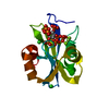 6wo7C  6wo8C  6wo9C 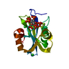 6wobC  6wocC  6wodC 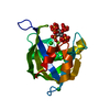 6woeC  6wofC 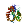 6wogC 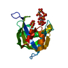 6wohC  6woiC C: citing same article ( |
|---|---|
| Similar structure data |


-Links
 PDBj
PDBj


- Assembly
| Deposited unit | 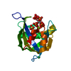
| ||||||||
|---|---|---|---|---|---|---|---|---|---|
| 1 |
| ||||||||
| Unit cell |
|
-Components
-Protein , 1 types, 1 molecules A
| #1: Protein | Mass: 19542.922 Da / Num. of mol.: 1 Source method: isolated from a genetically manipulated source Source: (gene. exp.) Homo sapiens (human) / Gene: NUDT3, DIPP, DIPP1 / Production host:  References: UniProt: O95989, diphosphoinositol-polyphosphate diphosphatase, Hydrolases; Acting on acid anhydrides; In phosphorus-containing anhydrides |
|---|
-Non-polymers , 6 types, 152 molecules 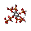


| #2: Chemical | ChemComp-UEV / ( Mass: 740.015 Da / Num. of mol.: 1 / Source method: obtained synthetically / Formula: C6H19O27P7 / Feature type: SUBJECT OF INVESTIGATION Mass: 740.015 Da / Num. of mol.: 1 / Source method: obtained synthetically / Formula: C6H19O27P7 / Feature type: SUBJECT OF INVESTIGATION | ||||||||
|---|---|---|---|---|---|---|---|---|---|
| #3: Chemical |  Mass: 24.305 Da / Num. of mol.: 3 / Source method: obtained synthetically / Formula: Mg / Feature type: SUBJECT OF INVESTIGATION Mass: 24.305 Da / Num. of mol.: 3 / Source method: obtained synthetically / Formula: Mg / Feature type: SUBJECT OF INVESTIGATION#4: Chemical | ChemComp-CL / |  Mass: 35.453 Da / Num. of mol.: 1 / Source method: obtained synthetically / Formula: Cl / Feature type: SUBJECT OF INVESTIGATION Mass: 35.453 Da / Num. of mol.: 1 / Source method: obtained synthetically / Formula: Cl / Feature type: SUBJECT OF INVESTIGATION#5: Chemical | ChemComp-SO4 / |  Mass: 96.063 Da / Num. of mol.: 1 / Source method: obtained synthetically / Formula: SO4 / Feature type: SUBJECT OF INVESTIGATION Mass: 96.063 Da / Num. of mol.: 1 / Source method: obtained synthetically / Formula: SO4 / Feature type: SUBJECT OF INVESTIGATION#6: Chemical | ChemComp-F / |  Mass: 18.998 Da / Num. of mol.: 1 / Source method: obtained synthetically / Formula: F / Feature type: SUBJECT OF INVESTIGATION Mass: 18.998 Da / Num. of mol.: 1 / Source method: obtained synthetically / Formula: F / Feature type: SUBJECT OF INVESTIGATION#7: Water | ChemComp-HOH / | Mass: 18.015 Da / Num. of mol.: 145 / Source method: isolated from a natural source / Formula: H2O |
-Details
| Has ligand of interest | Y |
|---|
-Experimental details
-Experiment
| Experiment | Method: X-RAY DIFFRACTION / Number of used crystals: 1 |
|---|
- Sample preparation
| Crystal | Density Matthews: 2.25 Å3/Da / Density % sol: 45.22 % |
|---|---|
| Crystal grow | Temperature: 293 K / Method: vapor diffusion, hanging drop / pH: 6 Details: 10% (w/v) PEG 8000, 10% (v/v) isopropanol, 200 mM Li2SO4, 75 mM NaAc, pH 5.5 and 25 mM HEPES, pH 7.0 and soaking in solution of 200 mM Li2SO4, 25% (w/v) PEG 8000, 10% (v/v) isopropanol, 75mM ...Details: 10% (w/v) PEG 8000, 10% (v/v) isopropanol, 200 mM Li2SO4, 75 mM NaAc, pH 5.5 and 25 mM HEPES, pH 7.0 and soaking in solution of 200 mM Li2SO4, 25% (w/v) PEG 8000, 10% (v/v) isopropanol, 75mM NaAc, pH 5.5, 25 mM HEPES, pH 7.0, 20 mM MgCl2 and 80 mM NaF in present of 4mM 2-IP7 PH range: 5.5-7.0 |
-Data collection
| Diffraction | Mean temperature: 100 K / Serial crystal experiment: N | |||||||||||||||||||||||||||||||||||||||||||||||||||||||||||||||||||||||||||||||||||||||||||||||||||||||||||||||||||||||||||||||||||||||||||||||||||||||||||||||||||||||||||||||||||||||||||||
|---|---|---|---|---|---|---|---|---|---|---|---|---|---|---|---|---|---|---|---|---|---|---|---|---|---|---|---|---|---|---|---|---|---|---|---|---|---|---|---|---|---|---|---|---|---|---|---|---|---|---|---|---|---|---|---|---|---|---|---|---|---|---|---|---|---|---|---|---|---|---|---|---|---|---|---|---|---|---|---|---|---|---|---|---|---|---|---|---|---|---|---|---|---|---|---|---|---|---|---|---|---|---|---|---|---|---|---|---|---|---|---|---|---|---|---|---|---|---|---|---|---|---|---|---|---|---|---|---|---|---|---|---|---|---|---|---|---|---|---|---|---|---|---|---|---|---|---|---|---|---|---|---|---|---|---|---|---|---|---|---|---|---|---|---|---|---|---|---|---|---|---|---|---|---|---|---|---|---|---|---|---|---|---|---|---|---|---|---|---|---|
| Diffraction source | Source: SYNCHROTRON / Site: APS / Beamline: 22-BM / Wavelength: 1 Å | |||||||||||||||||||||||||||||||||||||||||||||||||||||||||||||||||||||||||||||||||||||||||||||||||||||||||||||||||||||||||||||||||||||||||||||||||||||||||||||||||||||||||||||||||||||||||||||
| Detector | Type: RAYONIX MX300-HS / Detector: CCD / Date: Feb 16, 2020 | |||||||||||||||||||||||||||||||||||||||||||||||||||||||||||||||||||||||||||||||||||||||||||||||||||||||||||||||||||||||||||||||||||||||||||||||||||||||||||||||||||||||||||||||||||||||||||||
| Radiation | Protocol: SINGLE WAVELENGTH / Monochromatic (M) / Laue (L): M / Scattering type: x-ray | |||||||||||||||||||||||||||||||||||||||||||||||||||||||||||||||||||||||||||||||||||||||||||||||||||||||||||||||||||||||||||||||||||||||||||||||||||||||||||||||||||||||||||||||||||||||||||||
| Radiation wavelength | Wavelength: 1 Å / Relative weight: 1 | |||||||||||||||||||||||||||||||||||||||||||||||||||||||||||||||||||||||||||||||||||||||||||||||||||||||||||||||||||||||||||||||||||||||||||||||||||||||||||||||||||||||||||||||||||||||||||||
| Reflection | Resolution: 1.46→50 Å / Num. obs: 30051 / % possible obs: 96 % / Redundancy: 16.7 % / Rmerge(I) obs: 0.074 / Rpim(I) all: 0.017 / Rrim(I) all: 0.076 / Χ2: 0.981 / Net I/σ(I): 9.6 / Num. measured all: 500518 | |||||||||||||||||||||||||||||||||||||||||||||||||||||||||||||||||||||||||||||||||||||||||||||||||||||||||||||||||||||||||||||||||||||||||||||||||||||||||||||||||||||||||||||||||||||||||||||
| Reflection shell | Diffraction-ID: 1
|
- Processing
| Software |
| |||||||||||||||||||||||||||||||||||||||||||||||||||||||||||||||||
|---|---|---|---|---|---|---|---|---|---|---|---|---|---|---|---|---|---|---|---|---|---|---|---|---|---|---|---|---|---|---|---|---|---|---|---|---|---|---|---|---|---|---|---|---|---|---|---|---|---|---|---|---|---|---|---|---|---|---|---|---|---|---|---|---|---|---|
| Refinement | Method to determine structure: FOURIER SYNTHESIS / Resolution: 1.5→37.71 Å / Cor.coef. Fo:Fc: 0.972 / Cor.coef. Fo:Fc free: 0.97 / SU B: 2.109 / SU ML: 0.035 / Cross valid method: THROUGHOUT / σ(F): 0 / ESU R: 0.064 / ESU R Free: 0.053 / Stereochemistry target values: MAXIMUM LIKELIHOOD Details: HYDROGENS HAVE BEEN ADDED IN THE RIDING POSITIONS U VALUES : WITH TLS ADDED
| |||||||||||||||||||||||||||||||||||||||||||||||||||||||||||||||||
| Solvent computation | Ion probe radii: 0.8 Å / Shrinkage radii: 0.8 Å / VDW probe radii: 1.2 Å / Solvent model: MASK | |||||||||||||||||||||||||||||||||||||||||||||||||||||||||||||||||
| Displacement parameters | Biso max: 81.31 Å2 / Biso mean: 16.703 Å2 / Biso min: 7.5 Å2
| |||||||||||||||||||||||||||||||||||||||||||||||||||||||||||||||||
| Refinement step | Cycle: final / Resolution: 1.5→37.71 Å
| |||||||||||||||||||||||||||||||||||||||||||||||||||||||||||||||||
| Refine LS restraints |
| |||||||||||||||||||||||||||||||||||||||||||||||||||||||||||||||||
| LS refinement shell | Resolution: 1.5→1.539 Å / Rfactor Rfree error: 0 / Total num. of bins used: 20
| |||||||||||||||||||||||||||||||||||||||||||||||||||||||||||||||||
| Refinement TLS params. | Method: refined / Origin x: 6.209 Å / Origin y: 7.177 Å / Origin z: 13.827 Å
| |||||||||||||||||||||||||||||||||||||||||||||||||||||||||||||||||
| Refinement TLS group |
|
