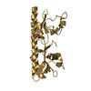
 Movie
Movie Controller


+ Open data

- Basic information
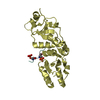
| Entry | Database: PDB / ID: 6vsi | ||||||
|---|---|---|---|---|---|---|---|
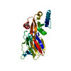
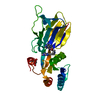
| Title | Crystal structure of FKBP12 of Candida auris | ||||||
 Components Components | Peptidylprolyl isomerase | ||||||
 Keywords Keywords | ISOMERASE / FKBP12 / C. auris / pathogenesis | ||||||
| Function / homology |  Function and homology information Function and homology informationpeptidylprolyl isomerase / peptidyl-prolyl cis-trans isomerase activity / cytoplasm Similarity search - Function | ||||||
| Biological species |  Candida auris (fungus) Candida auris (fungus) | ||||||
| Method |  X-RAY DIFFRACTION / SYNCHROTRON / MOLECULAR REPLACEMENT / molecular replacement / Resolution: 1.87 Å X-RAY DIFFRACTION / SYNCHROTRON / MOLECULAR REPLACEMENT / molecular replacement / Resolution: 1.87 Å | ||||||
 Authors Authors | Li, Z. / Li, H. / Hernandez, G. / LeMaster, D. | ||||||
| Funding support |  United States, 1items United States, 1items
| ||||||
 Citation Citation | Journal: Biochem.Biophys.Res.Commun. / Year: 2020 Title: Crystal structure and transient dimerization for the FKBP12 protein from the pathogenic fungus Candida auris. Authors: Bashir, Q. / Li, Z. / Li, H. / LeMaster, D.M. / Hernandez, G. | ||||||
| History |
|
- Structure visualization
| Structure viewer | Molecule: MolmilJmol/JSmol |
|---|
- Downloads & links
-Download
| PDBx/mmCIF format | 6vsi.cif.gz | 37.7 KB | Display | PDBx/mmCIF format |
|---|---|---|---|---|
| PDB format | pdb6vsi.ent.gz | 23.8 KB | Display | PDB format |
| PDBx/mmJSON format | 6vsi.json.gz | Tree view | PDBx/mmJSON format | |
| Others |  Other downloads Other downloads |
-Validation report
| Arichive directory | https://data.pdbj.org/pub/pdb/validation_reports/vs/6vsiftp://data.pdbj.org/pub/pdb/validation_reports/vs/6vsi | HTTPS FTP |
|---|
-Related structure data
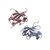
| Related structure data |  5ht1S S: Starting model for refinement |
|---|---|
| Similar structure data |


-Links
 PDBj
PDBj

- Assembly
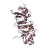
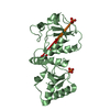
| Deposited unit | 
| |||||||||
|---|---|---|---|---|---|---|---|---|---|---|
| 1 | 
| |||||||||
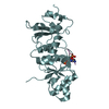
| Unit cell |
| |||||||||
| Components on special symmetry positions |
|
-Components
| #1: Protein | Mass: 11821.423 Da / Num. of mol.: 1 Source method: isolated from a genetically manipulated source Source: (gene. exp.) Candida auris (fungus) / Gene: CJJ09_002997 / Plasmid: pET11a / Production host:  References: UniProt: A0A5C1DY09, UniProt: A0A2H1A4Z6*PLUS, peptidylprolyl isomerase |
|---|---|
| #2: Chemical | ChemComp-SO4 /   Mass: 96.063 Da / Num. of mol.: 1 / Source method: obtained synthetically / Formula: SO4 Mass: 96.063 Da / Num. of mol.: 1 / Source method: obtained synthetically / Formula: SO4 |
| #3: Water | ChemComp-HOH /  Mass: 18.015 Da / Num. of mol.: 78 / Source method: isolated from a natural source / Formula: H2O Mass: 18.015 Da / Num. of mol.: 78 / Source method: isolated from a natural source / Formula: H2O |
| Has ligand of interest | N |
-Experimental details
-Experiment
| Experiment | Method: X-RAY DIFFRACTION / Number of used crystals: 1 |
|---|
- Sample preparation
| Crystal | Density Matthews: 4.05 Å3/Da / Density % sol: 70 % / Description: long rod |
|---|---|
| Crystal grow | Temperature: 298 K / Method: evaporation / pH: 7.5 Details: 2 uL 21.5 mg/mL protein in 20 mM Tris-HCl, pH 8.0, 200 mM sodium chloride + 2 uL 54% saturated ammonium sulfate, 0.1 M HEPES, pH 7.5, 2% isopropanol |
-Data collection
| Diffraction | Mean temperature: 100 K / Serial crystal experiment: N |
|---|---|
| Diffraction source | Source: SYNCHROTRON / Site: NSLS-II / Beamline: 19-ID / Wavelength: 0.9796 Å |
| Detector | Type: ADSC HF-4M / Detector: PIXEL / Date: Oct 30, 2019 |
| Radiation | Protocol: SINGLE WAVELENGTH / Monochromatic (M) / Laue (L): M / Scattering type: x-ray |
| Radiation wavelength | Wavelength: 0.9796 Å / Relative weight: 1 |
| Reflection | Resolution: 1.87→37.4 Å / Num. obs: 17303 / % possible obs: 91.89 % / Redundancy: 12.1 % / Biso Wilson estimate: 45.5 Å2 / CC1/2: 0.999 / CC star: 1 / Rmerge(I) obs: 0.071 / Rpim(I) all: 0.021 / Rrim(I) all: 0.074 / Net I/σ(I): 20.8 |
| Reflection shell | Resolution: 1.87→1.94 Å / Redundancy: 13.3 % / Num. unique obs: 571 / CC1/2: 0.29 / % possible all: 33.8 |
-Phasing
| Phasing | Method: molecular replacement | ||||||
|---|---|---|---|---|---|---|---|
| Phasing MR |
|
- Processing
| Software |
| ||||||||||||||||||||||||||||||||||||
|---|---|---|---|---|---|---|---|---|---|---|---|---|---|---|---|---|---|---|---|---|---|---|---|---|---|---|---|---|---|---|---|---|---|---|---|---|---|
| Refinement | Method to determine structure: MOLECULAR REPLACEMENT Starting model: PDB entry 5HT1 Resolution: 1.87→37.4 Å / SU ML: 0.2 / Cross valid method: THROUGHOUT / σ(F): 1.34 / Phase error: 29.16
| ||||||||||||||||||||||||||||||||||||
| Solvent computation | Shrinkage radii: 0.9 Å / VDW probe radii: 1.11 Å | ||||||||||||||||||||||||||||||||||||
| Displacement parameters | Biso max: 120.28 Å2 / Biso mean: 48.9185 Å2 / Biso min: 33.16 Å2 | ||||||||||||||||||||||||||||||||||||
| Refinement step | Cycle: final / Resolution: 1.87→37.4 Å
| ||||||||||||||||||||||||||||||||||||
| Refine LS restraints |
| ||||||||||||||||||||||||||||||||||||
| LS refinement shell | Refine-ID: X-RAY DIFFRACTION / Rfactor Rfree error: 0
|
