 Movie
Movie Controller

[English] 日本語
 Yorodumi
- PDB-6vpt: Crystal structure and mechanistic molecular modeling studies of R... -
+ Open data

- Basic information
| Entry | Database: PDB / ID: 6vpt | |||||||||
|---|---|---|---|---|---|---|---|---|---|---|
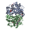
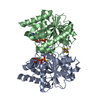
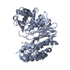
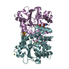
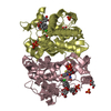
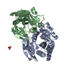
| Title | Crystal structure and mechanistic molecular modeling studies of Rv3377c: the Mycobacterium tuberculosis diterpene cyclase | |||||||||
 Components Components | Cyclase | |||||||||
 Keywords Keywords | LYASE / diterpene synthase / diterpene cyclase | |||||||||
| Function / homology |  Function and homology information Function and homology informationhalimadienyl-diphosphate synthase / geranylgeranyl diphosphate metabolic process / halimadienyl-diphosphate synthase activity / tuberculosinol biosynthetic process / diterpenoid biosynthetic process / cellular response to magnesium starvation / response to host immune response / terpene synthase activity / isomerase activity / lyase activity / magnesium ion binding Similarity search - Function | |||||||||
| Biological species |   Mycobacterium tuberculosis (bacteria) Mycobacterium tuberculosis (bacteria) | |||||||||
| Method |  X-RAY DIFFRACTION / SYNCHROTRON / MOLECULAR REPLACEMENT / Resolution: 2.718 Å X-RAY DIFFRACTION / SYNCHROTRON / MOLECULAR REPLACEMENT / Resolution: 2.718 Å | |||||||||
 Authors Authors | Zhang, T. / Prach, L. / DiMaio, F. / Siegel, J. | |||||||||
| Funding support |  United States, 2items United States, 2items
| |||||||||
 Citation Citation | Journal: Biochemistry / Year: 2020 Title: Crystal Structure and Mechanistic Molecular Modeling Studies of Mycobacterium tuberculosis Diterpene Cyclase Rv3377c. Authors: Zhang, Y. / Prach, L.M. / O'Brien, T.E. / DiMaio, F. / Prigozhin, D.M. / Corn, J.E. / Alber, T. / Siegel, J.B. / Tantillo, D.J. | |||||||||
| History |
|
- Structure visualization
| Structure viewer | Molecule: MolmilJmol/JSmol |
|---|
- Downloads & links
-Download
| PDBx/mmCIF format | 6vpt.cif.gz | 106.2 KB | Display | PDBx/mmCIF format |
|---|---|---|---|---|
| PDB format | pdb6vpt.ent.gz | 80.4 KB | Display | PDB format |
| PDBx/mmJSON format | 6vpt.json.gz | Tree view | PDBx/mmJSON format | |
| Others |  Other downloads Other downloads |
-Validation report
| Summary document | 6vpt_validation.pdf.gz | 247.2 KB | Display | wwPDB validaton report |
|---|---|---|---|---|
| Full document | 6vpt_full_validation.pdf.gz | 247.2 KB | Display | |
| Data in XML | 6vpt_validation.xml.gz | 1.1 KB | Display | |
| Data in CIF | 6vpt_validation.cif.gz | 5.6 KB | Display | |
| Arichive directory | https://data.pdbj.org/pub/pdb/validation_reports/vp/6vptftp://data.pdbj.org/pub/pdb/validation_reports/vp/6vpt | HTTPS FTP |
-Related structure data
| Related structure data |  3pyaS S: Starting model for refinement |
|---|---|
| Similar structure data |


-Links
 PDBj
PDBj

- Assembly
| Deposited unit | 
| ||||||||
|---|---|---|---|---|---|---|---|---|---|
| 1 |
| ||||||||
| Unit cell |
|
-Components
| #1: Protein | Mass: 55324.230 Da / Num. of mol.: 1 Source method: isolated from a genetically manipulated source Source: (gene. exp.) Mycobacterium tuberculosis (bacteria)Gene: ERS023446_02740, EZX46_08560, FDK60_02635, FDK62_05385, SAMEA2682864_03253, SAMEA2683035_03439 Production host: |
|---|---|
| #2: Water | ChemComp-HOH /  Mass: 18.015 Da / Num. of mol.: 32 / Source method: isolated from a natural source / Formula: H2O Mass: 18.015 Da / Num. of mol.: 32 / Source method: isolated from a natural source / Formula: H2O |
-Experimental details
-Experiment
| Experiment | Method: X-RAY DIFFRACTION / Number of used crystals: 1 |
|---|
- Sample preparation
| Crystal | Density Matthews: 2.24 Å3/Da / Density % sol: 45.16 % |
|---|---|
| Crystal grow | Temperature: 298 K / Method: vapor diffusion Details: 20% polyethylene glycol (PEG) 8000 and 0.1 M (cyclohexylamino) ethanesulfonic acid (CHES) pH 9.5 with 2 mM MgCl |
-Data collection
| Diffraction | Mean temperature: 298 K / Serial crystal experiment: N |
|---|---|
| Diffraction source | Source: SYNCHROTRON / Site: ALS / Beamline: 8.3.1 / Wavelength: 1 Å |
| Detector | Type: ADSC QUANTUM 315r / Detector: CCD / Date: Oct 1, 2007 |
| Radiation | Protocol: SINGLE WAVELENGTH / Monochromatic (M) / Laue (L): M / Scattering type: x-ray |
| Radiation wavelength | Wavelength: 1 Å / Relative weight: 1 |
| Reflection | Resolution: 2.718→50 Å / Num. obs: 12602 / % possible obs: 99.7 % / Redundancy: 3.9 % / Biso Wilson estimate: 47.05 Å2 / Rmerge(I) obs: 0.082 / Net I/σ(I): 15.8 |
| Reflection shell | Resolution: 2.718→2.99 Å / Rmerge(I) obs: 0.421 / Num. unique obs: 2974 / % possible all: 94 |
- Processing
| Software |
| ||||||||||||||||||||||||||||||
|---|---|---|---|---|---|---|---|---|---|---|---|---|---|---|---|---|---|---|---|---|---|---|---|---|---|---|---|---|---|---|---|
| Refinement | Method to determine structure: MOLECULAR REPLACEMENT Starting model: 3pya Resolution: 2.718→40.015 Å / SU ML: 0.36 / Cross valid method: THROUGHOUT / σ(F): 1.34 / Phase error: 24.46
| ||||||||||||||||||||||||||||||
| Solvent computation | Shrinkage radii: 0.9 Å / VDW probe radii: 1.11 Å | ||||||||||||||||||||||||||||||
| Displacement parameters | Biso max: 95.8 Å2 / Biso mean: 45.793 Å2 / Biso min: 18.36 Å2 | ||||||||||||||||||||||||||||||
| Refinement step | Cycle: final / Resolution: 2.718→40.015 Å
| ||||||||||||||||||||||||||||||
| LS refinement shell | Refine-ID: X-RAY DIFFRACTION / Rfactor Rfree error: 0
|
