| Entry | Database: PDB / ID: 6vpb
|
|---|
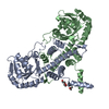
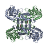
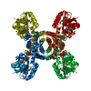
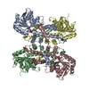
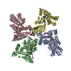
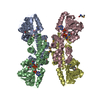
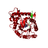
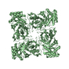
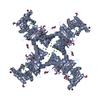
| Title | A novel membrane-bound 6-phosphogluconate dehydrogenase from the acetic acid bacteria Gluconacetobacter diazotrophicus (Gd6PGD) |
|---|
 Components Components | 6-phosphogluconate dehydrogenase |
|---|
 Keywords Keywords | OXIDOREDUCTASE / 6-phosphogluconate dehydrogenase / Gluconacetobacter diazotrophicus / membrane-bound protein / 6-phospho-D-gluconate / NAD+ |
|---|
| Function / homology |  Function and homology information Function and homology information
: / phosphogluconate dehydrogenase (decarboxylating) activity / D-gluconate metabolic process / pentose-phosphate shunt / NADP binding / metal ion bindingSimilarity search - Function 6-phosphogluconate dehydrogenase, YqeC-type / 6-phosphogluconate dehydrogenase, C-terminal / 6-phosphogluconate dehydrogenase / 6-phosphogluconate dehydrogenase, C-terminal domain / 6-phosphogluconate dehydrogenase, C-terminal domain / 6-phosphogluconate dehydrogenase, NADP-binding / NAD binding domain of 6-phosphogluconate dehydrogenase / 6-phosphogluconate dehydrogenase, domain 2 / 6-phosphogluconate dehydrogenase-like, C-terminal domain superfamily / NAD(P)-binding domain superfamilySimilarity search - Domain/homology |
|---|
| Biological species |  Gluconacetobacter diazotrophicus (bacteria) Gluconacetobacter diazotrophicus (bacteria) |
|---|
| Method |  X-RAY DIFFRACTION / SAD / Resolution: 1.87 Å X-RAY DIFFRACTION / SAD / Resolution: 1.87 Å |
|---|
 Authors Authors | Rodriguez-Romero, A. / Rodriguez-Hernandez, A. |
|---|
| Funding support |  Mexico, 1items Mexico, 1items | Organization | Grant number | Country |
|---|
| Consejo Nacional de Ciencia y Tecnologia (CONACYT) | 299048 | Mexico |
|
|---|
 Citation Citation | Journal: Febs J. / Year: 2021
Title: The structure of a novel membrane-associated 6-phosphogluconate dehydrogenase from Gluconacetobacter diazotrophicus (Gd6PGD) reveals a subfamily of short-chain 6PGDs.
Authors: Sarmiento-Pavia, P.D. / Rodriguez-Hernandez, A. / Rodriguez-Romero, A. / Sosa-Torres, M.E. |
|---|
| History | | Deposition | Feb 2, 2020 | Deposition site: RCSB / Processing site: RCSB |
|---|
| Revision 1.0 | Jul 22, 2020 | Provider: repository / Type: Initial release |
|---|
| Revision 1.1 | Mar 10, 2021 | Group: Database references / Category: citation
Item: _citation.journal_volume / _citation.page_first ..._citation.journal_volume / _citation.page_first / _citation.page_last / _citation.year |
|---|
| Revision 1.2 | Mar 6, 2024 | Group: Data collection / Database references / Refinement description
Category: chem_comp_atom / chem_comp_bond ...chem_comp_atom / chem_comp_bond / database_2 / struct_ncs_dom_lim
Item: _database_2.pdbx_DOI / _database_2.pdbx_database_accession ..._database_2.pdbx_DOI / _database_2.pdbx_database_accession / _struct_ncs_dom_lim.beg_auth_comp_id / _struct_ncs_dom_lim.beg_label_asym_id / _struct_ncs_dom_lim.beg_label_comp_id / _struct_ncs_dom_lim.beg_label_seq_id / _struct_ncs_dom_lim.end_auth_comp_id / _struct_ncs_dom_lim.end_label_asym_id / _struct_ncs_dom_lim.end_label_comp_id / _struct_ncs_dom_lim.end_label_seq_id |
|---|
|
|---|
 Movie
Movie Controller

 Yorodumi
 Open data
 Basic information
 Structure visualization
 Downloads & links
 Other downloads
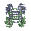
 PDBj
PDBj


 Assembly