 Movie
Movie Controller


+ Open data

- Basic information
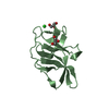
| Entry | Database: PDB / ID: 6vgv | ||||||
|---|---|---|---|---|---|---|---|
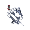
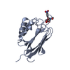
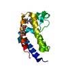
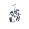
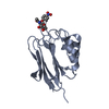
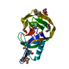
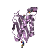
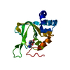
| Title | Crystal structure of VidaL intein | ||||||
 Components Components | VidaL | ||||||
 Keywords Keywords | SPLICING / Intein / split intein / atypical intein / protein splicing | ||||||
| Function / homology | Endonuclease - Pi-scei; Chain A, domain 1 / Hedgehog/Intein (Hint) domain / Beta Complex / Mainly Beta Function and homology information Function and homology information | ||||||
| Biological species | synthetic construct (others) | ||||||
| Method |  X-RAY DIFFRACTION / MOLECULAR REPLACEMENT / Resolution: 1.65 Å X-RAY DIFFRACTION / MOLECULAR REPLACEMENT / Resolution: 1.65 Å | ||||||
 Authors Authors | Burton, A.J. / Haugbro, M. / Parisi, E. / Muir, T.W. | ||||||
| Funding support |  United States, 1items United States, 1items
| ||||||
 Citation Citation | Journal: Proc.Natl.Acad.Sci.USA / Year: 2020 Title: Live-cell protein engineering with an ultra-short split intein. Authors: Burton, A.J. / Haugbro, M. / Parisi, E. / Muir, T.W. | ||||||
| History |
|
- Structure visualization
| Structure viewer | Molecule: MolmilJmol/JSmol |
|---|
- Downloads & links
-Download
| PDBx/mmCIF format | 6vgv.cif.gz | 88.4 KB | Display | PDBx/mmCIF format |
|---|---|---|---|---|
| PDB format | pdb6vgv.ent.gz | 55.2 KB | Display | PDB format |
| PDBx/mmJSON format | 6vgv.json.gz | Tree view | PDBx/mmJSON format | |
| Others |  Other downloads Other downloads |
-Validation report
| Arichive directory | https://data.pdbj.org/pub/pdb/validation_reports/vg/6vgvftp://data.pdbj.org/pub/pdb/validation_reports/vg/6vgv | HTTPS FTP |
|---|
-Related structure data


-Links
 PDBj
PDBj
- Assembly
| Deposited unit | 
| ||||||||||||
|---|---|---|---|---|---|---|---|---|---|---|---|---|---|
| 1 |
| ||||||||||||
| Unit cell |
|
-Components
| #1: Protein | Mass: 16179.247 Da / Num. of mol.: 1 / Mutation: C4A, N142A Source method: isolated from a genetically manipulated source Source: (gene. exp.) synthetic construct (others) / Production host:  | ||||
|---|---|---|---|---|---|
| #2: Chemical | ChemComp-GOL /   Mass: 92.094 Da / Num. of mol.: 6 / Source method: obtained synthetically / Formula: C3H8O3 Mass: 92.094 Da / Num. of mol.: 6 / Source method: obtained synthetically / Formula: C3H8O3#3: Water | ChemComp-HOH / |  Mass: 18.015 Da / Num. of mol.: 114 / Source method: isolated from a natural source / Formula: H2O Mass: 18.015 Da / Num. of mol.: 114 / Source method: isolated from a natural source / Formula: H2OHas ligand of interest | N | |
-Experimental details
-Experiment
| Experiment | Method: X-RAY DIFFRACTION / Number of used crystals: 1 |
|---|
- Sample preparation
| Crystal | Density Matthews: 2.22 Å3/Da / Density % sol: 44.67 % |
|---|---|
| Crystal grow | Temperature: 293 K / Method: vapor diffusion, sitting drop / pH: 4.6 Details: 0.1 M sodium acetate (pH 4.6) with 2 M ammonium sulfate |
-Data collection
| Diffraction | Mean temperature: 100 K / Serial crystal experiment: N |
|---|---|
| Diffraction source | Source: ROTATING ANODE / Type: RIGAKU MICROMAX-007 HF / Wavelength: 1.5418 Å |
| Detector | Type: DECTRIS PILATUS 300K / Detector: PIXEL / Date: Aug 30, 2018 |
| Radiation | Protocol: SINGLE WAVELENGTH / Monochromatic (M) / Laue (L): M / Scattering type: x-ray |
| Radiation wavelength | Wavelength: 1.5418 Å / Relative weight: 1 |
| Reflection | Resolution: 1.65→26.95 Å / Num. obs: 17134 / % possible obs: 98.9 % / Redundancy: 5.9 % / Biso Wilson estimate: 16.62 Å2 / CC1/2: 0.995 / Rpim(I) all: 0.023 / Rrim(I) all: 0.063 / Net I/σ(I): 4 |
| Reflection shell | Resolution: 1.65→1.75 Å / Mean I/σ(I) obs: 3.5 / Num. unique obs: 2567 / Rpim(I) all: 0.185 / Rrim(I) all: 0.323 / % possible all: 94.6 |
- Processing
| Software |
| ||||||||||||||||||||||||||||||||||||||||||||||||||||||||||||||||||||||||||||||||||||||||||||||||||||
|---|---|---|---|---|---|---|---|---|---|---|---|---|---|---|---|---|---|---|---|---|---|---|---|---|---|---|---|---|---|---|---|---|---|---|---|---|---|---|---|---|---|---|---|---|---|---|---|---|---|---|---|---|---|---|---|---|---|---|---|---|---|---|---|---|---|---|---|---|---|---|---|---|---|---|---|---|---|---|---|---|---|---|---|---|---|---|---|---|---|---|---|---|---|---|---|---|---|---|---|---|---|
| Refinement | Method to determine structure: MOLECULAR REPLACEMENT Starting model: VidaL SeMet structure Resolution: 1.65→26.95 Å / SU ML: 0.1386 / Cross valid method: FREE R-VALUE / σ(F): 1.36 / Phase error: 21.6494 / Stereochemistry target values: GeoStd + Monomer Library
| ||||||||||||||||||||||||||||||||||||||||||||||||||||||||||||||||||||||||||||||||||||||||||||||||||||
| Solvent computation | Shrinkage radii: 0.9 Å / VDW probe radii: 1.11 Å / Solvent model: FLAT BULK SOLVENT MODEL | ||||||||||||||||||||||||||||||||||||||||||||||||||||||||||||||||||||||||||||||||||||||||||||||||||||
| Displacement parameters | Biso mean: 21.75 Å2 | ||||||||||||||||||||||||||||||||||||||||||||||||||||||||||||||||||||||||||||||||||||||||||||||||||||
| Refinement step | Cycle: LAST / Resolution: 1.65→26.95 Å
| ||||||||||||||||||||||||||||||||||||||||||||||||||||||||||||||||||||||||||||||||||||||||||||||||||||
| Refine LS restraints |
| ||||||||||||||||||||||||||||||||||||||||||||||||||||||||||||||||||||||||||||||||||||||||||||||||||||
| LS refinement shell |
| ||||||||||||||||||||||||||||||||||||||||||||||||||||||||||||||||||||||||||||||||||||||||||||||||||||
| Refinement TLS params. | Method: refined / Refine-ID: X-RAY DIFFRACTION
| ||||||||||||||||||||||||||||||||||||||||||||||||||||||||||||||||||||||||||||||||||||||||||||||||||||
| Refinement TLS group |
|
