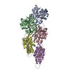
ムービー
ムービー コントローラー


+ データを開く

- 基本情報
基本情報
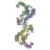
| 登録情報 | データベース: PDB / ID: 6vec | ||||||
|---|---|---|---|---|---|---|---|
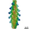
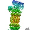
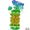
| タイトル | Cryo-EM structure of F-actin/Plastin2-ABD2 complex | ||||||
 要素 要素 |
| ||||||
 キーワード キーワード | PROTEIN FIBRIL / F-actin / Plastin 2 / Helical reconstruction | ||||||
| 機能・相同性 |  機能・相同性情報 機能・相同性情報actin filament network formation / actin crosslink formation / T cell activation involved in immune response / : / cortical actin cytoskeleton organization / cytoskeletal motor activator activity / regulation of intracellular protein transport / myosin heavy chain binding / tropomyosin binding / actin filament bundle ...actin filament network formation / actin crosslink formation / T cell activation involved in immune response / : / cortical actin cytoskeleton organization / cytoskeletal motor activator activity / regulation of intracellular protein transport / myosin heavy chain binding / tropomyosin binding / actin filament bundle / troponin I binding / filamentous actin / mesenchyme migration / skeletal muscle myofibril / actin filament bundle assembly / striated muscle thin filament / skeletal muscle thin filament assembly / animal organ regeneration / actin monomer binding / glial cell projection / phagocytic cup / skeletal muscle fiber development / ruffle / stress fiber / titin binding / actin filament polymerization / Gene and protein expression by JAK-STAT signaling after Interleukin-12 stimulation / filopodium / actin filament / integrin binding / 加水分解酵素; 酸無水物に作用; 酸無水物に作用・細胞または細胞小器官の運動に関与 / ruffle membrane / calcium-dependent protein binding / actin filament binding / cell junction / cell migration / lamellipodium / actin cytoskeleton / actin binding / cell body / GTPase binding / protein domain specific binding / focal adhesion / hydrolase activity / calcium ion binding / positive regulation of gene expression / perinuclear region of cytoplasm / magnesium ion binding / : / extracellular exosome / ATP binding / identical protein binding / plasma membrane / cytoplasm / cytosol 類似検索 - 分子機能 | ||||||
| 生物種 |  Homo sapiens (ヒト) Homo sapiens (ヒト) | ||||||
| 手法 | 電子顕微鏡法 / らせん対称体再構成法 / クライオ電子顕微鏡法 / 解像度: 3.9 Å | ||||||
 データ登録者 データ登録者 | Zheng, W. / Kudryashov, D.S. / Egelman, E.H. | ||||||
| 資金援助 |  米国, 1件 米国, 1件
| ||||||
 引用 引用 | ジャーナル: Bone Res / 年: 2020 タイトル: Osteogenesis imperfecta mutations in plastin 3 lead to impaired calcium regulation of actin bundling. 著者: Christopher L Schwebach / Elena Kudryashova / Weili Zheng / Matthew Orchard / Harper Smith / Lucas A Runyan / Edward H Egelman / Dmitri S Kudryashov / 要旨: Mutations in actin-bundling protein plastin 3 (PLS3) emerged as a cause of congenital osteoporosis, but neither the role of PLS3 in bone development nor the mechanisms underlying PLS3-dependent ...Mutations in actin-bundling protein plastin 3 (PLS3) emerged as a cause of congenital osteoporosis, but neither the role of PLS3 in bone development nor the mechanisms underlying PLS3-dependent osteoporosis are understood. Of the over 20 identified osteoporosis-linked PLS3 mutations, we investigated all five that are expected to produce full-length protein. One of the mutations distorted an actin-binding loop in the second actin-binding domain of PLS3 and abolished F-actin bundling as revealed by cryo-EM reconstruction and protein interaction assays. Surprisingly, the remaining four mutants fully retained F-actin bundling ability. However, they displayed defects in Ca sensitivity: two of the mutants lost the ability to be inhibited by Ca, while the other two became hypersensitive to Ca. Each group of the mutants with similar biochemical properties showed highly characteristic cellular behavior. Wild-type PLS3 was distributed between lamellipodia and focal adhesions. In striking contrast, the Ca-hyposensitive mutants were not found at the leading edge but localized exclusively at focal adhesions/stress fibers, which displayed reinforced morphology. Consistently, the Ca-hypersensitive PLS3 mutants were restricted to lamellipodia, while chelation of Ca caused their redistribution to focal adhesions. Finally, the bundling-deficient mutant failed to co-localize with any F-actin structures in cells despite a preserved F-actin binding through a non-mutation-bearing actin-binding domain. Our findings revealed that severe osteoporosis can be caused by a mutational disruption of the Ca-controlled PLS3's cycling between adhesion complexes and the leading edge. Integration of the structural, biochemical, and cell biology insights enabled us to propose a molecular mechanism of plastin activity regulation by Ca. | ||||||
| 履歴 |
|
- 構造の表示
構造の表示
| ムービー |
ムービービューア |
|---|---|
| 構造ビューア | 分子: MolmilJmol/JSmol |
- ダウンロードとリンク
-ダウンロード
| PDBx/mmCIF形式 | 6vec.cif.gz | 1.1 MB | 表示 | PDBx/mmCIF形式 |
|---|---|---|---|---|
| PDB形式 | pdb6vec.ent.gz | 954.3 KB | 表示 | PDB形式 |
| PDBx/mmJSON形式 | 6vec.json.gz | ツリー表示 | PDBx/mmJSON形式 | |
| その他 |  その他のダウンロード その他のダウンロード |
-検証レポート
| アーカイブディレクトリ | https://data.pdbj.org/pub/pdb/validation_reports/ve/6vecftp://data.pdbj.org/pub/pdb/validation_reports/ve/6vec | HTTPS FTP |
|---|
-関連構造データ


-リンク
 PDBj
PDBj


- 集合体
集合体
| 登録構造単位 | 
|
|---|---|
| 1 |
|
| 対称性 | らせん対称: (回転対称性: 1 / Dyad axis: no / N subunits divisor: 1 / Num. of operations: 11 / Rise per n subunits: 28 Å / Rotation per n subunits: -166.5 °) |
-要素
| #1: タンパク質 | 分子量: 42096.953 Da / 分子数: 11 / 由来タイプ: 天然 / 由来: (天然) #2: タンパク質 | 分子量: 47983.633 Da / 分子数: 11 / Fragment: UNP residues 385-625 / 由来タイプ: 組換発現 / 由来: (組換発現) Homo sapiens (ヒト) / 遺伝子: HEL-S-37発現宿主:  参照: UniProt: V9HWJ7, UniProt: P13796*PLUS #3: 化合物 | ChemComp-ADP /   分子量: 427.201 Da / 分子数: 11 / 由来タイプ: 合成 / 式: C10H15N5O10P2 / コメント: ADP, エネルギー貯蔵分子*YM 分子量: 427.201 Da / 分子数: 11 / 由来タイプ: 合成 / 式: C10H15N5O10P2 / コメント: ADP, エネルギー貯蔵分子*YM#4: 化合物 | ChemComp-MG /   分子量: 24.305 Da / 分子数: 11 / 由来タイプ: 合成 / 式: Mg 分子量: 24.305 Da / 分子数: 11 / 由来タイプ: 合成 / 式: Mg研究の焦点であるリガンドがあるか | N | |
|---|
-実験情報
-実験
| 実験 | 手法: 電子顕微鏡法 |
|---|---|
| EM実験 | 試料の集合状態: FILAMENT / 3次元再構成法: らせん対称体再構成法 |
- 試料調製
試料調製
| 構成要素 |
| ||||||||||||||||||||||||
|---|---|---|---|---|---|---|---|---|---|---|---|---|---|---|---|---|---|---|---|---|---|---|---|---|---|
| 分子量 | 実験値: NO | ||||||||||||||||||||||||
| 由来(天然) |
| ||||||||||||||||||||||||
| 由来(組換発現) | 生物種: | ||||||||||||||||||||||||
| 緩衝液 | pH: 7.5 | ||||||||||||||||||||||||
| 試料 | 包埋: NO / シャドウイング: NO / 染色: NO / 凍結: YES | ||||||||||||||||||||||||
| 試料支持 | 詳細: unspecified | ||||||||||||||||||||||||
| 急速凍結 | 凍結剤: ETHANE |
- 電子顕微鏡撮影
| 実験機器 |  モデル: Titan Krios / 画像提供: FEI Company |
|---|---|
| 顕微鏡 | モデル: FEI TITAN KRIOS |
| 電子銃 | 電子線源:  FIELD EMISSION GUN / 加速電圧: 300 kV / 照射モード: FLOOD BEAM FIELD EMISSION GUN / 加速電圧: 300 kV / 照射モード: FLOOD BEAM |
| 電子レンズ | モード: BRIGHT FIELD |
| 試料ホルダ | 凍結剤: NITROGEN |
| 撮影 | 電子線照射量: 20 e/Å2 / 検出モード: INTEGRATING フィルム・検出器のモデル: FEI FALCON III (4k x 4k) |
- 解析
解析
| EMソフトウェア |
| ||||||||||||||||||||||||||||||||||||
|---|---|---|---|---|---|---|---|---|---|---|---|---|---|---|---|---|---|---|---|---|---|---|---|---|---|---|---|---|---|---|---|---|---|---|---|---|---|
| CTF補正 | タイプ: PHASE FLIPPING AND AMPLITUDE CORRECTION | ||||||||||||||||||||||||||||||||||||
| らせん対称 | 回転角度/サブユニット: -166.5 ° / 軸方向距離/サブユニット: 28 Å / らせん対称軸の対称性: C1 | ||||||||||||||||||||||||||||||||||||
| 粒子像の選択 | 選択した粒子像数: 248184 | ||||||||||||||||||||||||||||||||||||
| 3次元再構成 | 解像度: 3.9 Å / 解像度の算出法: FSC 0.143 CUT-OFF / 粒子像の数: 124092 / 対称性のタイプ: HELICAL | ||||||||||||||||||||||||||||||||||||
| 原子モデル構築 | PDB-ID: 5ONV Accession code: 5ONV / Source name: PDB / タイプ: experimental model |