 Movie
Movie Controller

[English] 日本語
 Yorodumi
- PDB-6ve1: Crystal structure of endo-beta-N-acetylglucosaminidase H at high pH -
+ Open data

- Basic information
| Entry | Database: PDB / ID: 6ve1 | ||||||
|---|---|---|---|---|---|---|---|
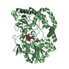
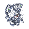
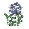
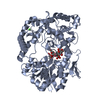
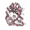
| Title | Crystal structure of endo-beta-N-acetylglucosaminidase H at high pH | ||||||
 Components Components | Endo-beta-N-acetylglucosaminidase H | ||||||
 Keywords Keywords | HYDROLASE / SUGAR BINDING PROTEIN / enzyme / deglycosylase / post-translational modification | ||||||
| Function / homology |  Function and homology information Function and homology informationmannosyl-glycoprotein endo-beta-N-acetylglucosaminidase / mannosyl-glycoprotein endo-beta-N-acetylglucosaminidase activity / carbohydrate metabolic process / extracellular region Similarity search - Function | ||||||
| Biological species |  Streptomyces plicatus (bacteria) Streptomyces plicatus (bacteria) | ||||||
| Method |  X-RAY DIFFRACTION / SYNCHROTRON / MOLECULAR REPLACEMENT / Resolution: 2.1 Å X-RAY DIFFRACTION / SYNCHROTRON / MOLECULAR REPLACEMENT / Resolution: 2.1 Å | ||||||
 Authors Authors | Stachowski, T.R. / Snell, M.E. / Snell, E.S. | ||||||
| Funding support |  United States, 1items United States, 1items
| ||||||
 Citation Citation | Journal: Plos One / Year: 2020 Title: SAXS studies of X-ray induced disulfide bond damage: Engineering high-resolution insight from a low-resolution technique. Authors: Stachowski, T.R. / Snell, M.E. / Snell, E.H. | ||||||
| History |
|
- Structure visualization
| Structure viewer | Molecule: MolmilJmol/JSmol |
|---|
- Downloads & links
-Download
| PDBx/mmCIF format | 6ve1.cif.gz | 399.9 KB | Display | PDBx/mmCIF format |
|---|---|---|---|---|
| PDB format | pdb6ve1.ent.gz | 328.1 KB | Display | PDB format |
| PDBx/mmJSON format | 6ve1.json.gz | Tree view | PDBx/mmJSON format | |
| Others |  Other downloads Other downloads |
-Validation report
| Arichive directory | https://data.pdbj.org/pub/pdb/validation_reports/ve/6ve1ftp://data.pdbj.org/pub/pdb/validation_reports/ve/6ve1 | HTTPS FTP |
|---|
-Related structure data
| Related structure data |  1edtS S: Starting model for refinement |
|---|---|
| Similar structure data |


-Links
 PDBj
PDBj- Assembly
| Deposited unit | 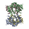
| ||||||||
|---|---|---|---|---|---|---|---|---|---|
| 1 | 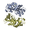
| ||||||||
| 2 | 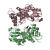
| ||||||||
| Unit cell |
|
-Components
| #1: Protein | Mass: 30393.506 Da / Num. of mol.: 4 / Fragment: UNP residues 47-313 Source method: isolated from a genetically manipulated source Source: (gene. exp.) Streptomyces plicatus (bacteria) / Production host: References: UniProt: P04067, mannosyl-glycoprotein endo-beta-N-acetylglucosaminidase #2: Chemical | ChemComp-MG /   Mass: 24.305 Da / Num. of mol.: 6 / Source method: obtained synthetically / Formula: Mg Mass: 24.305 Da / Num. of mol.: 6 / Source method: obtained synthetically / Formula: Mg#3: Water | ChemComp-HOH / |  Mass: 18.015 Da / Num. of mol.: 1136 / Source method: isolated from a natural source / Formula: H2O Mass: 18.015 Da / Num. of mol.: 1136 / Source method: isolated from a natural source / Formula: H2OHas ligand of interest | N | |
|---|
-Experimental details
-Experiment
| Experiment | Method: X-RAY DIFFRACTION / Number of used crystals: 1 |
|---|
- Sample preparation
| Crystal | Density Matthews: 2.58 Å3/Da / Density % sol: 52.4 % |
|---|---|
| Crystal grow | Temperature: 295 K / Method: batch mode / pH: 9 / Details: PEG20000, magnesium nitrate, TAPS, pH 9.0 |
-Data collection
| Diffraction | Mean temperature: 100 K / Serial crystal experiment: N |
|---|---|
| Diffraction source | Source: SYNCHROTRON / Site: APS / Beamline: 17-ID / Wavelength: 1 Å |
| Detector | Type: DECTRIS PILATUS 6M / Detector: PIXEL / Date: Dec 15, 2019 |
| Radiation | Protocol: SINGLE WAVELENGTH / Monochromatic (M) / Laue (L): M / Scattering type: x-ray |
| Radiation wavelength | Wavelength: 1 Å / Relative weight: 1 |
| Reflection | Resolution: 2.1→41.031 Å / Num. obs: 74108 / % possible obs: 99.69 % / Redundancy: 2 % / CC1/2: 0.986 / Net I/σ(I): 5.89 |
| Reflection shell | Resolution: 2.1→2.175 Å / Num. unique obs: 7330 / CC1/2: 0.618 |
- Processing
| Software |
| ||||||||||||||||||||||||||||||||||||||||||||||||||||||||||||||||||||||||||||||||||||||||||||||||||||||||||||||||||||||||||||||||||||||||||||||||||||||||||||||||||
|---|---|---|---|---|---|---|---|---|---|---|---|---|---|---|---|---|---|---|---|---|---|---|---|---|---|---|---|---|---|---|---|---|---|---|---|---|---|---|---|---|---|---|---|---|---|---|---|---|---|---|---|---|---|---|---|---|---|---|---|---|---|---|---|---|---|---|---|---|---|---|---|---|---|---|---|---|---|---|---|---|---|---|---|---|---|---|---|---|---|---|---|---|---|---|---|---|---|---|---|---|---|---|---|---|---|---|---|---|---|---|---|---|---|---|---|---|---|---|---|---|---|---|---|---|---|---|---|---|---|---|---|---|---|---|---|---|---|---|---|---|---|---|---|---|---|---|---|---|---|---|---|---|---|---|---|---|---|---|---|---|---|---|---|
| Refinement | Method to determine structure: MOLECULAR REPLACEMENT Starting model: PDB entry 1EDT Resolution: 2.1→41.031 Å / SU ML: 0.28 / Cross valid method: THROUGHOUT / σ(F): 1.97 / Phase error: 31.22
| ||||||||||||||||||||||||||||||||||||||||||||||||||||||||||||||||||||||||||||||||||||||||||||||||||||||||||||||||||||||||||||||||||||||||||||||||||||||||||||||||||
| Solvent computation | Shrinkage radii: 0.9 Å / VDW probe radii: 1.11 Å | ||||||||||||||||||||||||||||||||||||||||||||||||||||||||||||||||||||||||||||||||||||||||||||||||||||||||||||||||||||||||||||||||||||||||||||||||||||||||||||||||||
| Displacement parameters | Biso max: 70.5 Å2 / Biso mean: 16.786 Å2 / Biso min: 1.82 Å2 | ||||||||||||||||||||||||||||||||||||||||||||||||||||||||||||||||||||||||||||||||||||||||||||||||||||||||||||||||||||||||||||||||||||||||||||||||||||||||||||||||||
| Refinement step | Cycle: final / Resolution: 2.1→41.031 Å
| ||||||||||||||||||||||||||||||||||||||||||||||||||||||||||||||||||||||||||||||||||||||||||||||||||||||||||||||||||||||||||||||||||||||||||||||||||||||||||||||||||
| LS refinement shell | Refine-ID: X-RAY DIFFRACTION / Rfactor Rfree error: 0
|
