 Movie
Movie Controller

[English] 日本語
 Yorodumi
- PDB-6vdh: Crystal structure of ancestral apicomplexan lactate dehydrogenase... -
+ Open data

- Basic information
| Entry | Database: PDB / ID: 6vdh | ||||||
|---|---|---|---|---|---|---|---|
| Title | Crystal structure of ancestral apicomplexan lactate dehydrogenase in alternate dimer configuration with sulfate. | ||||||
 Components Components | lactate dehydrogenase | ||||||
 Keywords Keywords | OXIDOREDUCTASE / Nucleotide Binding / Rossman Fold / Ancestral Reconstruction | ||||||
| Function / homology | L-2-Hydroxyisocaproate Dehydrogenase; Chain A, domain 2 / Lactate dehydrogenase/glycoside hydrolase, family 4, C-terminal / NAD(P)-binding Rossmann-like Domain / Alpha-Beta Complex / Rossmann fold / 3-Layer(aba) Sandwich / Alpha Beta Function and homology information Function and homology information | ||||||
| Biological species |  Apicomplexa sp. (eukaryote) Apicomplexa sp. (eukaryote) | ||||||
| Method |  X-RAY DIFFRACTION / SYNCHROTRON / MOLECULAR REPLACEMENT / Resolution: 1.85 Å X-RAY DIFFRACTION / SYNCHROTRON / MOLECULAR REPLACEMENT / Resolution: 1.85 Å | ||||||
 Authors Authors | Theobald, D.L. / Wirth, J.D. / Classen, S. / Perlmutter, N. | ||||||
| Funding support |  United States, 1items United States, 1items
| ||||||
 Citation Citation | Journal: To Be Published Title: Mechanism of Bifunctionality in an Ancestral Apicomplexan Malate/Lactate Dehydrogenase Authors: Theobald, D.L. / Wirth, J.D. / Classen, S. / Perlmutter, N. | ||||||
| History |
|
- Structure visualization
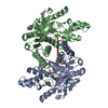
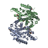
| Structure viewer | Molecule: MolmilJmol/JSmol |
|---|
- Downloads & links
-Download
| PDBx/mmCIF format | 6vdh.cif.gz | 272.3 KB | Display | PDBx/mmCIF format |
|---|---|---|---|---|
| PDB format | pdb6vdh.ent.gz | 179.2 KB | Display | PDB format |
| PDBx/mmJSON format | 6vdh.json.gz | Tree view | PDBx/mmJSON format | |
| Others |  Other downloads Other downloads |
-Validation report
| Arichive directory | https://data.pdbj.org/pub/pdb/validation_reports/vd/6vdhftp://data.pdbj.org/pub/pdb/validation_reports/vd/6vdh | HTTPS FTP |
|---|
-Related structure data
| Related structure data |  6vdiC  6vdjC  4plgS S: Starting model for refinement C: citing same article ( |
|---|---|
| Similar structure data |


-Links
 PDBj
PDBj- Assembly
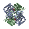
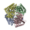
| Deposited unit | 
| ||||||||||||
|---|---|---|---|---|---|---|---|---|---|---|---|---|---|
| 1 | 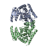
| ||||||||||||
| Unit cell |
|
-Components
| #1: Protein | Mass: 35905.477 Da / Num. of mol.: 2 Source method: isolated from a genetically manipulated source Details: Ancestral apicomplexan lactate dehydrogenase in alternate dimer conformation with sulfate bound Source: (gene. exp.) Apicomplexa sp. (eukaryote) / Plasmid: pET24a(+) / Production host:  #2: Chemical |   Mass: 96.063 Da / Num. of mol.: 2 / Source method: obtained synthetically / Formula: SO4 / Feature type: SUBJECT OF INVESTIGATION Mass: 96.063 Da / Num. of mol.: 2 / Source method: obtained synthetically / Formula: SO4 / Feature type: SUBJECT OF INVESTIGATION#3: Water | ChemComp-HOH / |  Mass: 18.015 Da / Num. of mol.: 323 / Source method: isolated from a natural source / Formula: H2O Mass: 18.015 Da / Num. of mol.: 323 / Source method: isolated from a natural source / Formula: H2OHas ligand of interest | Y | |
|---|
-Experimental details
-Experiment
| Experiment | Method: X-RAY DIFFRACTION / Number of used crystals: 1 |
|---|
- Sample preparation
| Crystal | Density Matthews: 2.78 Å3/Da / Density % sol: 55.8 % |
|---|---|
| Crystal grow | Temperature: 293 K / Method: vapor diffusion, hanging drop Details: 20mg/mL protein, 2mM NADH4; well solution: 5% v/v 2-Propanol, 2.5M Ammonium Sulfate |
-Data collection
| Diffraction | Mean temperature: 90 K / Serial crystal experiment: N |
|---|---|
| Diffraction source | Source: SYNCHROTRON / Site: ALS / Beamline: 12.3.1 / Wavelength: 1.115866 Å |
| Detector | Type: ADSC QUANTUM 315r / Detector: CCD / Date: Oct 16, 2019 |
| Radiation | Monochromator: Si(111) / Protocol: SINGLE WAVELENGTH / Monochromatic (M) / Laue (L): M / Scattering type: x-ray |
| Radiation wavelength | Wavelength: 1.115866 Å / Relative weight: 1 |
| Reflection | Resolution: 1.68→45.53 Å / Num. obs: 86980 / % possible obs: 88.9 % / Redundancy: 5.92 % / Biso Wilson estimate: 30.34 Å2 / CC1/2: 0.998 / Rrim(I) all: 0.102 / Χ2: 0.86 / Net I/σ(I): 3.66 |
| Reflection shell | Resolution: 1.68→1.79 Å / Redundancy: 3.18 % / Mean I/σ(I) obs: 0.18 / Num. unique obs: 18335 / CC1/2: 0.071 / Rrim(I) all: 5.295 / % possible all: 62.6 |
- Processing
| Software |
| ||||||||||||||||||||||||||||||||||||||||||||||||||||||||||||||||||||||||||||||||||||
|---|---|---|---|---|---|---|---|---|---|---|---|---|---|---|---|---|---|---|---|---|---|---|---|---|---|---|---|---|---|---|---|---|---|---|---|---|---|---|---|---|---|---|---|---|---|---|---|---|---|---|---|---|---|---|---|---|---|---|---|---|---|---|---|---|---|---|---|---|---|---|---|---|---|---|---|---|---|---|---|---|---|---|---|---|---|
| Refinement | Method to determine structure: MOLECULAR REPLACEMENT Starting model: 4PLG Resolution: 1.85→45.53 Å / SU ML: 0.3457 / Cross valid method: FREE R-VALUE / σ(F): 1.33 / Phase error: 32.0699 Stereochemistry target values: GeoStd + Monomer Library + CDL v1.2
| ||||||||||||||||||||||||||||||||||||||||||||||||||||||||||||||||||||||||||||||||||||
| Solvent computation | Shrinkage radii: 0.9 Å / VDW probe radii: 1.11 Å / Solvent model: FLAT BULK SOLVENT MODEL | ||||||||||||||||||||||||||||||||||||||||||||||||||||||||||||||||||||||||||||||||||||
| Displacement parameters | Biso mean: 40.49 Å2 | ||||||||||||||||||||||||||||||||||||||||||||||||||||||||||||||||||||||||||||||||||||
| Refinement step | Cycle: LAST / Resolution: 1.85→45.53 Å
| ||||||||||||||||||||||||||||||||||||||||||||||||||||||||||||||||||||||||||||||||||||
| Refine LS restraints |
| ||||||||||||||||||||||||||||||||||||||||||||||||||||||||||||||||||||||||||||||||||||
| LS refinement shell |
|
