 Movie
Movie Controller


+ Open data

- Basic information
| Entry | Database: PDB / ID: 6vco | ||||||
|---|---|---|---|---|---|---|---|
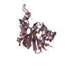
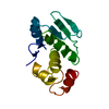
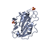
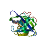
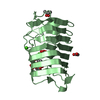
| Title | Crystal structure of E.coli RppH in complex with ppcpA | ||||||
 Components Components | RNA pyrophosphohydrolase | ||||||
 Keywords Keywords | RNA BINDING PROTEIN / RNA degradation | ||||||
| Function / homology |  Function and homology information Function and homology informationRNA NAD-cap (NMN-forming) hydrolase activity / RNA destabilization / mRNA 5'-diphosphatase activity / RNA decapping / NAD-cap decapping / hydrolase activity, acting on acid anhydrides, in phosphorus-containing anhydrides / tRNA processing / mRNA catabolic process / Hydrolases; Acting on acid anhydrides; In phosphorus-containing anhydrides / magnesium ion binding / cytoplasm Similarity search - Function | ||||||
| Biological species |  | ||||||
| Method |  X-RAY DIFFRACTION / MOLECULAR REPLACEMENT / Resolution: 1.7 Å X-RAY DIFFRACTION / MOLECULAR REPLACEMENT / Resolution: 1.7 Å | ||||||
 Authors Authors | Gao, A. / Vasilyev, N. / Kaushik, A. / Duan, W. / Serganov, A. | ||||||
| Funding support |  United States, 1items United States, 1items
| ||||||
 Citation Citation | Journal: Nucleic Acids Res. / Year: 2020 Title: Principles of RNA and nucleotide discrimination by the RNA processing enzyme RppH. Authors: Gao, A. / Vasilyev, N. / Kaushik, A. / Duan, W. / Serganov, A. | ||||||
| History |
|
- Structure visualization
| Structure viewer | Molecule: MolmilJmol/JSmol |
|---|
- Downloads & links
-Download
| PDBx/mmCIF format | 6vco.cif.gz | 82.3 KB | Display | PDBx/mmCIF format |
|---|---|---|---|---|
| PDB format | pdb6vco.ent.gz | 58.9 KB | Display | PDB format |
| PDBx/mmJSON format | 6vco.json.gz | Tree view | PDBx/mmJSON format | |
| Others |  Other downloads Other downloads |
-Validation report
| Arichive directory | https://data.pdbj.org/pub/pdb/validation_reports/vc/6vcoftp://data.pdbj.org/pub/pdb/validation_reports/vc/6vco | HTTPS FTP |
|---|
-Related structure data
| Related structure data |  6vckC 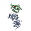 6vclC 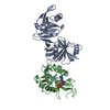 6vcmC 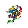 6vcnC 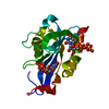 6vcpC  6vcqC 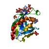 6vcrC 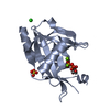 4s2yS C: citing same article ( S: Starting model for refinement |
|---|---|
| Similar structure data |


-Links
 PDBj
PDBj


- Assembly
| Deposited unit | 
| ||||||||||||
|---|---|---|---|---|---|---|---|---|---|---|---|---|---|
| 1 |
| ||||||||||||
| Unit cell |
|
-Components
| #1: Protein | Mass: 18965.773 Da / Num. of mol.: 1 / Mutation: Q159A, E160A Source method: isolated from a genetically manipulated source Source: (gene. exp.) Strain: K12 / Gene: rppH, nudH, ygdP, b2830, JW2798 / Production host: References: UniProt: P0A776, Hydrolases; Acting on acid anhydrides; In phosphorus-containing anhydrides |
|---|---|
| #2: Chemical | ChemComp-APC /   Mass: 505.208 Da / Num. of mol.: 1 / Source method: obtained synthetically / Formula: C11H18N5O12P3 / Feature type: SUBJECT OF INVESTIGATION / Comment: AMP-CPP, energy-carrying molecule analogue*YM Mass: 505.208 Da / Num. of mol.: 1 / Source method: obtained synthetically / Formula: C11H18N5O12P3 / Feature type: SUBJECT OF INVESTIGATION / Comment: AMP-CPP, energy-carrying molecule analogue*YM |
| #3: Water | ChemComp-HOH /  Mass: 18.015 Da / Num. of mol.: 153 / Source method: isolated from a natural source / Formula: H2O Mass: 18.015 Da / Num. of mol.: 153 / Source method: isolated from a natural source / Formula: H2O |
| Has ligand of interest | Y |
-Experimental details
-Experiment
| Experiment | Method: X-RAY DIFFRACTION / Number of used crystals: 1 |
|---|
- Sample preparation
| Crystal | Density Matthews: 2.34 Å3/Da / Density % sol: 47.36 % |
|---|---|
| Crystal grow | Temperature: 293.15 K / Method: vapor diffusion, hanging drop Details: 0.2 M (NH4)2SO4, 6.25% (v/v) PEG3350, and 5% (v/v) glycerol |
-Data collection
| Diffraction | Mean temperature: 100 K / Serial crystal experiment: N |
|---|---|
| Diffraction source | Source: ROTATING ANODE / Type: RIGAKU MICROMAX-007 / Wavelength: 1.5418 Å |
| Detector | Type: RIGAKU RAXIS IV++ / Detector: IMAGE PLATE / Date: May 16, 2013 |
| Radiation | Protocol: SINGLE WAVELENGTH / Monochromatic (M) / Laue (L): M / Scattering type: x-ray |
| Radiation wavelength | Wavelength: 1.5418 Å / Relative weight: 1 |
| Reflection | Resolution: 1.7→19.48 Å / Num. obs: 18406 / % possible obs: 95.3 % / Redundancy: 7.1 % / Biso Wilson estimate: 22.06 Å2 / CC1/2: 0.999 / Net I/σ(I): 24 |
| Reflection shell | Resolution: 1.7→1.76 Å / Num. unique obs: 1706 / CC1/2: 0.923 |
- Processing
| Software |
| ||||||||||||||||||||||||||||||||||||||||||||||||||||||||
|---|---|---|---|---|---|---|---|---|---|---|---|---|---|---|---|---|---|---|---|---|---|---|---|---|---|---|---|---|---|---|---|---|---|---|---|---|---|---|---|---|---|---|---|---|---|---|---|---|---|---|---|---|---|---|---|---|---|
| Refinement | Method to determine structure: MOLECULAR REPLACEMENT Starting model: 4S2Y Resolution: 1.7→19.48 Å / SU ML: 0.1848 / Cross valid method: FREE R-VALUE / σ(F): 1.36 / Phase error: 22.7444
| ||||||||||||||||||||||||||||||||||||||||||||||||||||||||
| Solvent computation | Shrinkage radii: 0.9 Å / VDW probe radii: 1.11 Å | ||||||||||||||||||||||||||||||||||||||||||||||||||||||||
| Displacement parameters | Biso mean: 31.57 Å2 | ||||||||||||||||||||||||||||||||||||||||||||||||||||||||
| Refinement step | Cycle: LAST / Resolution: 1.7→19.48 Å
| ||||||||||||||||||||||||||||||||||||||||||||||||||||||||
| Refine LS restraints |
| ||||||||||||||||||||||||||||||||||||||||||||||||||||||||
| LS refinement shell |
|
