 Movie
Movie Controller


+ Open data

- Basic information
| Entry | Database: PDB / ID: 6v4v | ||||||
|---|---|---|---|---|---|---|---|
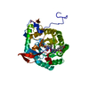
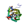
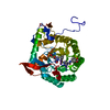
| Title | The crystal structure of BonA from Acinetobacter baumannii | ||||||
 Components Components | BON domain protein | ||||||
 Keywords Keywords | LIPID BINDING PROTEIN / Periplasmic / Lipoprotein / Divisome Protein / Cell Motility / Outer-membrane stability | ||||||
| Function / homology | : / BON domain profile. / BON domain / BON domain / Prokaryotic membrane lipoprotein lipid attachment site profile. / metal ion binding / BON domain-containing protein Function and homology information Function and homology information | ||||||
| Biological species |  Acinetobacter baumannii (bacteria) Acinetobacter baumannii (bacteria) | ||||||
| Method |  X-RAY DIFFRACTION / SYNCHROTRON / SAD / Resolution: 1.65 Å X-RAY DIFFRACTION / SYNCHROTRON / SAD / Resolution: 1.65 Å | ||||||
 Authors Authors | Grinter, R. | ||||||
| Funding support |  Australia, 1items Australia, 1items
| ||||||
 Citation Citation | Journal: Mbio / Year: 2021 Title: BonA from Acinetobacter baumannii Forms a Divisome-Localized Decamer That Supports Outer Envelope Function. Authors: Grinter, R. / Morris, F.C. / Dunstan, R.A. / Leung, P.M. / Kropp, A. / Belousoff, M. / Gunasinghe, S.D. / Scott, N.E. / Beckham, S. / Peleg, A.Y. / Greening, C. / Li, J. / Heinz, E. / Lithgow, T. | ||||||
| History |
|
- Structure visualization
| Structure viewer | Molecule: MolmilJmol/JSmol |
|---|
- Downloads & links
-Download
| PDBx/mmCIF format | 6v4v.cif.gz | 79.9 KB | Display | PDBx/mmCIF format |
|---|---|---|---|---|
| PDB format | pdb6v4v.ent.gz | 59 KB | Display | PDB format |
| PDBx/mmJSON format | 6v4v.json.gz | Tree view | PDBx/mmJSON format | |
| Others |  Other downloads Other downloads |
-Validation report
| Arichive directory | https://data.pdbj.org/pub/pdb/validation_reports/v4/6v4vftp://data.pdbj.org/pub/pdb/validation_reports/v4/6v4v | HTTPS FTP |
|---|
-Related structure data
| Similar structure data |
|---|


-Links
 PDBj
PDBj


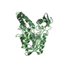
- Assembly
| Deposited unit | 
| ||||||||||||
|---|---|---|---|---|---|---|---|---|---|---|---|---|---|
| 1 | 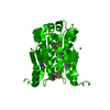
| ||||||||||||
| Unit cell |
| ||||||||||||
| Components on special symmetry positions |
|
-Components
| #1: Protein | Mass: 20646.668 Da / Num. of mol.: 1 Source method: isolated from a genetically manipulated source Source: (gene. exp.) Acinetobacter baumannii (bacteria) / Gene: A7M79_12275 / Production host: | ||||||
|---|---|---|---|---|---|---|---|
| #2: Chemical | ChemComp-ZN /   Mass: 65.409 Da / Num. of mol.: 4 / Source method: obtained synthetically / Formula: Zn Mass: 65.409 Da / Num. of mol.: 4 / Source method: obtained synthetically / Formula: Zn#3: Water | ChemComp-HOH / |  Mass: 18.015 Da / Num. of mol.: 153 / Source method: isolated from a natural source / Formula: H2O Mass: 18.015 Da / Num. of mol.: 153 / Source method: isolated from a natural source / Formula: H2OHas ligand of interest | N | Has protein modification | Y | |
-Experimental details
-Experiment
| Experiment | Method: X-RAY DIFFRACTION / Number of used crystals: 1 |
|---|
- Sample preparation
| Crystal | Density Matthews: 2.53 Å3/Da / Density % sol: 51.29 % |
|---|---|
| Crystal grow | Temperature: 298 K / Method: vapor diffusion, sitting drop / pH: 4.5 / Details: 0.2 M Zn Acetate, 0.1 M Na Acetate, 20 % PEG 3350 |
-Data collection
| Diffraction | Mean temperature: 100 K / Serial crystal experiment: N |
|---|---|
| Diffraction source | Source: SYNCHROTRON / Site: Australian Synchrotron / Beamline: MX1 / Wavelength: 0.987 Å |
| Detector | Type: MAR CCD 130 mm / Detector: CCD / Date: Mar 17, 2016 / Details: Yes |
| Radiation | Protocol: SINGLE WAVELENGTH / Monochromatic (M) / Laue (L): M / Scattering type: x-ray |
| Radiation wavelength | Wavelength: 0.987 Å / Relative weight: 1 |
| Reflection | Resolution: 1.65→46.07 Å / Num. obs: 25589 / % possible obs: 99.9 % / Redundancy: 10.7 % / CC1/2: 1 / Rmerge(I) obs: 0.057 / Rpim(I) all: 0.023 / Net I/σ(I): 24.1 |
| Reflection shell | Resolution: 1.65→1.68 Å / Redundancy: 10.8 % / Rmerge(I) obs: 1.675 / Num. unique obs: 1254 / CC1/2: 0.693 / Rpim(I) all: 0.777 / % possible all: 100 |
- Processing
| Software |
| ||||||||||||||||||||||||||||||||||||||||
|---|---|---|---|---|---|---|---|---|---|---|---|---|---|---|---|---|---|---|---|---|---|---|---|---|---|---|---|---|---|---|---|---|---|---|---|---|---|---|---|---|---|
| Refinement | Method to determine structure: SAD / Resolution: 1.65→35.904 Å / SU ML: 0.19 / Cross valid method: THROUGHOUT / σ(F): 1.34 / Phase error: 23.48
| ||||||||||||||||||||||||||||||||||||||||
| Solvent computation | Shrinkage radii: 0.9 Å / VDW probe radii: 1.11 Å | ||||||||||||||||||||||||||||||||||||||||
| Displacement parameters | Biso max: 86.69 Å2 / Biso mean: 38.1331 Å2 / Biso min: 18.6 Å2 | ||||||||||||||||||||||||||||||||||||||||
| Refinement step | Cycle: final / Resolution: 1.65→35.904 Å
| ||||||||||||||||||||||||||||||||||||||||
| Refinement TLS params. | Method: refined / Origin x: 9.2099 Å / Origin y: 47.0654 Å / Origin z: 39.8639 Å
| ||||||||||||||||||||||||||||||||||||||||
| Refinement TLS group |
|
