 Movie
Movie Controller

[English] 日本語
 Yorodumi
- PDB-6usm: Structure of nuclease domain of human parvovirus B19 non-structur... -
+ Open data

- Basic information
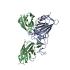
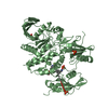
| Entry | Database: PDB / ID: 6usm | ||||||
|---|---|---|---|---|---|---|---|
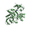
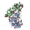
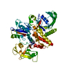
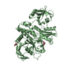
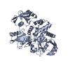
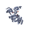
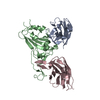
| Title | Structure of nuclease domain of human parvovirus B19 non-structural protein 1 in complex with zinc | ||||||
 Components Components |
| ||||||
 Keywords Keywords | REPLICATION / B19 / DNA binding protein | ||||||
| Function / homology |  Function and homology information Function and homology informationdetection of maltose stimulus / maltose transport complex / carbohydrate transport / carbohydrate transmembrane transporter activity / maltose binding / maltose transport / maltodextrin transmembrane transport / ATP-binding cassette (ABC) transporter complex, substrate-binding subunit-containing / ATP-binding cassette (ABC) transporter complex / viral genome replication ...detection of maltose stimulus / maltose transport complex / carbohydrate transport / carbohydrate transmembrane transporter activity / maltose binding / maltose transport / maltodextrin transmembrane transport / ATP-binding cassette (ABC) transporter complex, substrate-binding subunit-containing / ATP-binding cassette (ABC) transporter complex / viral genome replication / cell chemotaxis / outer membrane-bounded periplasmic space / endonuclease activity / periplasmic space / DNA replication / DNA damage response / host cell nucleus / DNA binding / ATP binding / membrane / metal ion binding Similarity search - Function | ||||||
| Biological species |   Human parvovirus B19 Human parvovirus B19 | ||||||
| Method |  X-RAY DIFFRACTION / SYNCHROTRON / MOLECULAR REPLACEMENT / Resolution: 3.37 Å X-RAY DIFFRACTION / SYNCHROTRON / MOLECULAR REPLACEMENT / Resolution: 3.37 Å | ||||||
 Authors Authors | Tewary, S.K. | ||||||
 Citation Citation | Journal: To Be Published Title: B19 Parvovirus Non-structural protein 1 in complex with zinc Authors: Tewary, S.K. | ||||||
| History |
|
- Structure visualization
| Structure viewer | Molecule: MolmilJmol/JSmol |
|---|
- Downloads & links
-Download
| PDBx/mmCIF format | 6usm.cif.gz | 118.5 KB | Display | PDBx/mmCIF format |
|---|---|---|---|---|
| PDB format | pdb6usm.ent.gz | 88.9 KB | Display | PDB format |
| PDBx/mmJSON format | 6usm.json.gz | Tree view | PDBx/mmJSON format | |
| Others |  Other downloads Other downloads |
-Validation report
| Arichive directory | https://data.pdbj.org/pub/pdb/validation_reports/us/6usmftp://data.pdbj.org/pub/pdb/validation_reports/us/6usm | HTTPS FTP |
|---|
-Related structure data
| Similar structure data |
|---|
-Links
 PDBj
PDBj


- Assembly
| Deposited unit | 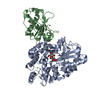
| ||||||||||
|---|---|---|---|---|---|---|---|---|---|---|---|
| 1 |
| ||||||||||
| Unit cell |
|
-Components
| #1: Protein | Mass: 40805.289 Da / Num. of mol.: 1 Source method: isolated from a genetically manipulated source Source: (gene. exp.) |
|---|---|
| #2: Protein | Mass: 19475.391 Da / Num. of mol.: 1 Source method: isolated from a genetically manipulated source Source: (gene. exp.) Human parvovirus B19 / Gene: NS / Production host: Expression vector pET-mod (others) / References: UniProt: Q6TV13 |
| #3: Polysaccharide | alpha-D-glucopyranose-(1-4)-alpha-D-glucopyranose / alpha-maltose  Source method: isolated from a genetically manipulated source Details: oligosaccharide / References: alpha-maltose |
| #4: Chemical | ChemComp-ZN /   Mass: 65.409 Da / Num. of mol.: 1 / Source method: obtained synthetically / Formula: Zn / Feature type: SUBJECT OF INVESTIGATION Mass: 65.409 Da / Num. of mol.: 1 / Source method: obtained synthetically / Formula: Zn / Feature type: SUBJECT OF INVESTIGATION |
| Has ligand of interest | Y |
-Experimental details
-Experiment
| Experiment | Method: X-RAY DIFFRACTION / Number of used crystals: 1 |
|---|
- Sample preparation
| Crystal | Density Matthews: 3.43 Å3/Da / Density % sol: 64.18 % |
|---|---|
| Crystal grow | Temperature: 293 K / Method: vapor diffusion, hanging drop / pH: 4.6 Details: 1.2 M LiSo4 and sodium acetate tri-hydrate buffer of pH4.6 |
-Data collection
| Diffraction | Mean temperature: 273 K / Serial crystal experiment: N |
|---|---|
| Diffraction source | Source: SYNCHROTRON / Site: APS  / Beamline: 23-ID-B / Wavelength: 1.033 Å / Beamline: 23-ID-B / Wavelength: 1.033 Å |
| Detector | Type: DECTRIS EIGER X 16M / Detector: PIXEL / Date: Sep 3, 2013 |
| Radiation | Protocol: SINGLE WAVELENGTH / Monochromatic (M) / Laue (L): M / Scattering type: x-ray |
| Radiation wavelength | Wavelength: 1.033 Å / Relative weight: 1 |
| Reflection | Resolution: 3.37→47.955 Å / Num. obs: 11564 / % possible obs: 96.8 % / Redundancy: 3 % / Biso Wilson estimate: 81.22 Å2 / Rmerge(I) obs: 0.125 / Net I/σ(I): 8.7 |
| Reflection shell | Resolution: 3.37→48 Å / Num. unique obs: 11564 / Rsym value: 0.125 |
- Processing
| Software |
| ||||||||||||||||||||||||
|---|---|---|---|---|---|---|---|---|---|---|---|---|---|---|---|---|---|---|---|---|---|---|---|---|---|
| Refinement | Method to determine structure: MOLECULAR REPLACEMENT Starting model: MBP Resolution: 3.37→47 Å / Cross valid method: THROUGHOUT /
| ||||||||||||||||||||||||
| Displacement parameters | Biso mean: 88.23 Å2 | ||||||||||||||||||||||||
| Refinement step | Cycle: LAST / Resolution: 3.37→47 Å
| ||||||||||||||||||||||||
| Refine LS restraints |
|
