 Movie
Movie Controller

[English] 日本語
 Yorodumi
- PDB-6ufe: The structure of a potassium selective ion channel at atomic reso... -
+ Open data

- Basic information
| Entry | Database: PDB / ID: 6ufe | ||||||
|---|---|---|---|---|---|---|---|
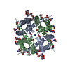
| Title | The structure of a potassium selective ion channel at atomic resolution | ||||||
 Components Components | Transporter | ||||||
 Keywords Keywords | MEMBRANE PROTEIN / ion channel / potassium ion channel | ||||||
| Function / homology |  Function and homology information Function and homology informationpotassium channel activity / membrane / metal ion binding / identical protein binding Similarity search - Function | ||||||
| Biological species |  | ||||||
| Method |  X-RAY DIFFRACTION / SYNCHROTRON / MOLECULAR REPLACEMENT / Resolution: 1.2 Å X-RAY DIFFRACTION / SYNCHROTRON / MOLECULAR REPLACEMENT / Resolution: 1.2 Å | ||||||
 Authors Authors | Langan, P.S. / Vandavasi, V.G. / Sullivan, B. / Afonine, P.V. / Weiss, K.L. | ||||||
 Citation Citation | Journal: Iucrj / Year: 2020 Title: The structure of a potassium-selective ion channel reveals a hydrophobic gate regulating ion permeation. Authors: Langan, P.S. / Vandavasi, V.G. / Kopec, W. / Sullivan, B. / Afonne, P.V. / Weiss, K.L. / de Groot, B.L. / Coates, L. | ||||||
| History |
|
- Structure visualization
| Structure viewer | Molecule: MolmilJmol/JSmol |
|---|
- Downloads & links
-Download
| PDBx/mmCIF format | 6ufe.cif.gz | 133.7 KB | Display | PDBx/mmCIF format |
|---|---|---|---|---|
| PDB format | pdb6ufe.ent.gz | 108.3 KB | Display | PDB format |
| PDBx/mmJSON format | 6ufe.json.gz | Tree view | PDBx/mmJSON format | |
| Others |  Other downloads Other downloads |
-Validation report
| Arichive directory | https://data.pdbj.org/pub/pdb/validation_reports/uf/6ufeftp://data.pdbj.org/pub/pdb/validation_reports/uf/6ufe | HTTPS FTP |
|---|
-Related structure data
| Related structure data |  3oufS S: Starting model for refinement |
|---|---|
| Similar structure data |


-Links
 PDBj
PDBj

- Assembly
| Deposited unit | 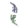
| |||||||||||||||||||||||||||
|---|---|---|---|---|---|---|---|---|---|---|---|---|---|---|---|---|---|---|---|---|---|---|---|---|---|---|---|---|
| 1 | 
| |||||||||||||||||||||||||||
| 2 | 
| |||||||||||||||||||||||||||
| Unit cell |
| |||||||||||||||||||||||||||
| Components on special symmetry positions |
|
-Components
| #1: Protein | Mass: 10195.952 Da / Num. of mol.: 2 / Mutation: D66Y, N68D Source method: isolated from a genetically manipulated source Source: (gene. exp.) Gene: A9485_19160, BACERE00184_02078, CJ306_03585, CN950_06075, CN980_22870, COI98_17615, CON37_12595 Production host: #2: Chemical | ChemComp-MPD / (   Mass: 118.174 Da / Num. of mol.: 10 / Source method: obtained synthetically / Formula: C6H14O2 / Comment: precipitant*YM Mass: 118.174 Da / Num. of mol.: 10 / Source method: obtained synthetically / Formula: C6H14O2 / Comment: precipitant*YM#3: Chemical | ChemComp-K /   Mass: 39.098 Da / Num. of mol.: 8 / Source method: obtained synthetically / Formula: K Mass: 39.098 Da / Num. of mol.: 8 / Source method: obtained synthetically / Formula: K#4: Water | ChemComp-HOH / |  Mass: 18.015 Da / Num. of mol.: 83 / Source method: isolated from a natural source / Formula: H2O Mass: 18.015 Da / Num. of mol.: 83 / Source method: isolated from a natural source / Formula: H2OHas ligand of interest | N | |
|---|
-Experimental details
-Experiment
| Experiment | Method: X-RAY DIFFRACTION / Number of used crystals: 1 |
|---|
- Sample preparation
| Crystal | Density Matthews: 2.53 Å3/Da / Density % sol: 51.31 % |
|---|---|
| Crystal grow | Temperature: 293 K / Method: vapor diffusion, sitting drop / Details: See paper for details |
-Data collection
| Diffraction | Mean temperature: 100 K / Serial crystal experiment: N |
|---|---|
| Diffraction source | Source: SYNCHROTRON / Site: APS  / Beamline: 19-ID / Wavelength: 0.97 Å / Beamline: 19-ID / Wavelength: 0.97 Å |
| Detector | Type: ADSC QUANTUM 315r / Detector: CCD / Date: Jun 6, 2017 |
| Radiation | Protocol: SINGLE WAVELENGTH / Monochromatic (M) / Laue (L): M / Scattering type: x-ray |
| Radiation wavelength | Wavelength: 0.97 Å / Relative weight: 1 |
| Reflection | Resolution: 1.2→30 Å / Num. obs: 61537 / % possible obs: 97.4 % / Redundancy: 5.34 % / CC1/2: 0.998 / Net I/σ(I): 12.23 |
| Reflection shell | Resolution: 1.2→1.23 Å / Num. unique obs: 3673 / CC1/2: 0.489 |
- Processing
| Software |
| ||||||||||||||||||||||||
|---|---|---|---|---|---|---|---|---|---|---|---|---|---|---|---|---|---|---|---|---|---|---|---|---|---|
| Refinement | Method to determine structure: MOLECULAR REPLACEMENT Starting model: 3OUF Resolution: 1.2→21.459 Å / SU ML: 0.1 / Cross valid method: FREE R-VALUE / σ(F): 1.34 / Phase error: 14.76 / Stereochemistry target values: ML
| ||||||||||||||||||||||||
| Solvent computation | Shrinkage radii: 0.9 Å / VDW probe radii: 1.11 Å / Solvent model: FLAT BULK SOLVENT MODEL | ||||||||||||||||||||||||
| Refinement step | Cycle: LAST / Resolution: 1.2→21.459 Å
| ||||||||||||||||||||||||
| Refine LS restraints |
| ||||||||||||||||||||||||
| LS refinement shell | Resolution: 1.2→1.23 Å
|
