 Movie
Movie Controller

[English] 日本語
 Yorodumi
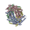
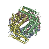
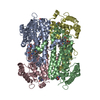
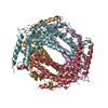
- PDB-6ud5: Crystal structure of human tryptophan 2,3-dioxygenase in complex ... -
+ Open data

- Basic information
| Entry | Database: PDB / ID: 6ud5 | |||||||||
|---|---|---|---|---|---|---|---|---|---|---|
| Title | Crystal structure of human tryptophan 2,3-dioxygenase in complex with carbon monoxide and tryptophan | |||||||||
 Components Components | Tryptophan 2,3-dioxygenase | |||||||||
 Keywords Keywords | OXIDOREDUCTASE / Tryptophan Dioxygenase / carbon monoxide / Tryptophan | |||||||||
| Function / homology |  Function and homology information Function and homology informationresponse to nitroglycerin / L-tryptophan catabolic process to acetyl-CoA / tryptophan 2,3-dioxygenase / tryptophan 2,3-dioxygenase activity / L-tryptophan catabolic process to kynurenine / Tryptophan catabolism / amino acid binding / oxygen binding / protein homotetramerization / heme binding ...response to nitroglycerin / L-tryptophan catabolic process to acetyl-CoA / tryptophan 2,3-dioxygenase / tryptophan 2,3-dioxygenase activity / L-tryptophan catabolic process to kynurenine / Tryptophan catabolism / amino acid binding / oxygen binding / protein homotetramerization / heme binding / metal ion binding / identical protein binding / cytosol Similarity search - Function | |||||||||
| Biological species |  Homo sapiens (human) Homo sapiens (human) | |||||||||
| Method |  X-RAY DIFFRACTION / SYNCHROTRON / MOLECULAR REPLACEMENT / Resolution: 2.05 Å X-RAY DIFFRACTION / SYNCHROTRON / MOLECULAR REPLACEMENT / Resolution: 2.05 Å | |||||||||
 Authors Authors | Pham, K.N. / Lewis-Ballester, A. / Yeh, S.R. | |||||||||
| Funding support |  United States, 2items United States, 2items
| |||||||||
 Citation Citation | Journal: J.Am.Chem.Soc. / Year: 2021 Title: Conformational Plasticity in Human Heme-Based Dioxygenases. Authors: Pham, K.N. / Lewis-Ballester, A. / Yeh, S.R. | |||||||||
| History |
|
- Structure visualization
| Structure viewer | Molecule: MolmilJmol/JSmol |
|---|
- Downloads & links
-Download
| PDBx/mmCIF format | 6ud5.cif.gz | 321.9 KB | Display | PDBx/mmCIF format |
|---|---|---|---|---|
| PDB format | pdb6ud5.ent.gz | 258 KB | Display | PDB format |
| PDBx/mmJSON format | 6ud5.json.gz | Tree view | PDBx/mmJSON format | |
| Others |  Other downloads Other downloads |
-Validation report
| Arichive directory | https://data.pdbj.org/pub/pdb/validation_reports/ud/6ud5ftp://data.pdbj.org/pub/pdb/validation_reports/ud/6ud5 | HTTPS FTP |
|---|
-Related structure data
| Related structure data |  6ubpC 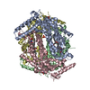 5ti9S C: citing same article ( S: Starting model for refinement |
|---|---|
| Similar structure data |


-Links
 PDBj
PDBj


- Assembly
| Deposited unit | 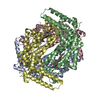
| ||||||||
|---|---|---|---|---|---|---|---|---|---|
| 1 |
| ||||||||
| Unit cell |
|
-Components
| #1: Protein | Mass: 45182.535 Da / Num. of mol.: 4 / Fragment: UNP residues 18-389 Source method: isolated from a genetically manipulated source Source: (gene. exp.) Homo sapiens (human) / Gene: TDO2, TDO / Production host:  #2: Chemical | ChemComp-HEM /   Mass: 616.487 Da / Num. of mol.: 4 / Source method: obtained synthetically / Formula: C34H32FeN4O4 Mass: 616.487 Da / Num. of mol.: 4 / Source method: obtained synthetically / Formula: C34H32FeN4O4#3: Chemical | ChemComp-CMO /   Mass: 28.010 Da / Num. of mol.: 4 / Source method: obtained synthetically / Formula: CO Mass: 28.010 Da / Num. of mol.: 4 / Source method: obtained synthetically / Formula: CO#4: Chemical | ChemComp-TRP /   Type: L-peptide linking / Mass: 204.225 Da / Num. of mol.: 8 / Source method: obtained synthetically / Formula: C11H12N2O2 / Feature type: SUBJECT OF INVESTIGATION Type: L-peptide linking / Mass: 204.225 Da / Num. of mol.: 8 / Source method: obtained synthetically / Formula: C11H12N2O2 / Feature type: SUBJECT OF INVESTIGATION#5: Water | ChemComp-HOH / |  Mass: 18.015 Da / Num. of mol.: 773 / Source method: isolated from a natural source / Formula: H2O Mass: 18.015 Da / Num. of mol.: 773 / Source method: isolated from a natural source / Formula: H2OHas ligand of interest | Y | |
|---|
-Experimental details
-Experiment
| Experiment | Method: X-RAY DIFFRACTION / Number of used crystals: 1 |
|---|
- Sample preparation
| Crystal | Density Matthews: 2.97 Å3/Da / Density % sol: 58.56 % |
|---|---|
| Crystal grow | Temperature: 298 K / Method: microbatch / pH: 5 Details: 50 mM sodium citrate, pH 5.6, 2% Tacsimate, pH 5.0, 5% PEG3350 PH range: 5.6 |
-Data collection
| Diffraction | Mean temperature: 100 K / Serial crystal experiment: N |
|---|---|
| Diffraction source | Source: SYNCHROTRON / Site: APS / Beamline: 31-ID / Wavelength: 0.97931 Å |
| Detector | Type: RAYONIX MX325HE / Detector: CCD / Date: Apr 8, 2016 |
| Radiation | Monochromator: diamond(111) / Protocol: SINGLE WAVELENGTH / Monochromatic (M) / Laue (L): M / Scattering type: x-ray |
| Radiation wavelength | Wavelength: 0.97931 Å / Relative weight: 1 |
| Reflection | Resolution: 2.05→19.97 Å / Num. obs: 120121 / % possible obs: 96.7 % / Redundancy: 6.7 % / CC1/2: 0.99 / Net I/σ(I): 7.9 |
| Reflection shell | Resolution: 2.05→2.16 Å / Mean I/σ(I) obs: 1.9 / Num. unique obs: 16664 / CC1/2: 0.59 |
- Processing
| Software |
| ||||||||||||||||||||||||||||||||||||||||||||||||||||||||||||
|---|---|---|---|---|---|---|---|---|---|---|---|---|---|---|---|---|---|---|---|---|---|---|---|---|---|---|---|---|---|---|---|---|---|---|---|---|---|---|---|---|---|---|---|---|---|---|---|---|---|---|---|---|---|---|---|---|---|---|---|---|---|
| Refinement | Method to determine structure: MOLECULAR REPLACEMENT Starting model: PDB entry 5TI9 Resolution: 2.05→19.97 Å / Cor.coef. Fo:Fc: 0.962 / Cor.coef. Fo:Fc free: 0.945 / SU B: 4.865 / SU ML: 0.124 / Cross valid method: THROUGHOUT / σ(F): 0 / ESU R: 0.173 / ESU R Free: 0.153 Details: HYDROGENS HAVE BEEN ADDED IN THE RIDING POSITIONS U VALUES : REFINED INDIVIDUALLY
| ||||||||||||||||||||||||||||||||||||||||||||||||||||||||||||
| Solvent computation | Ion probe radii: 0.8 Å / Shrinkage radii: 0.8 Å / VDW probe radii: 1.2 Å | ||||||||||||||||||||||||||||||||||||||||||||||||||||||||||||
| Displacement parameters | Biso max: 128.59 Å2 / Biso mean: 37.501 Å2 / Biso min: 20.85 Å2
| ||||||||||||||||||||||||||||||||||||||||||||||||||||||||||||
| Refinement step | Cycle: final / Resolution: 2.05→19.97 Å
| ||||||||||||||||||||||||||||||||||||||||||||||||||||||||||||
| Refine LS restraints |
| ||||||||||||||||||||||||||||||||||||||||||||||||||||||||||||
| LS refinement shell | Resolution: 2.05→2.103 Å / Rfactor Rfree error: 0 / Total num. of bins used: 20
|
