National Institutes of Health/National Human Genome Research Institute (NIH/NHGRI)
R01-HL12565
United States
National Institutes of Health/National Human Genome Research Institute (NIH/NHGRI)
R35-GM122510
United States
Citation
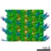
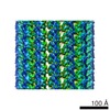
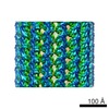
Journal: Proc Natl Acad Sci U S A / Year: 2020 Title: The structure of helical lipoprotein lipase reveals an unexpected twist in lipase storage. Authors: Kathryn H Gunn / Benjamin S Roberts / Fengbin Wang / Joshua D Strauss / Mario J Borgnia / Edward H Egelman / Saskia B Neher / Abstract: Lipases are enzymes necessary for the proper distribution and utilization of lipids in the human body. Lipoprotein lipase (LPL) is active in capillaries, where it plays a crucial role in preventing ...Lipases are enzymes necessary for the proper distribution and utilization of lipids in the human body. Lipoprotein lipase (LPL) is active in capillaries, where it plays a crucial role in preventing dyslipidemia by hydrolyzing triglycerides from packaged lipoproteins. Thirty years ago, the existence of a condensed and inactive LPL oligomer was proposed. Although recent work has shed light on the structure of the LPL monomer, the inactive oligomer remained opaque. Here we present a cryo-EM reconstruction of a helical LPL oligomer at 3.8-Å resolution. Helix formation is concentration-dependent, and helices are composed of inactive dihedral LPL dimers. Heparin binding stabilizes LPL helices, and the presence of substrate triggers helix disassembly. Superresolution fluorescent microscopy of endogenous LPL revealed that LPL adopts a filament-like distribution in vesicles. Mutation of one of the helical LPL interaction interfaces causes loss of the filament-like distribution. Taken together, this suggests that LPL is condensed into its inactive helical form for storage in intracellular vesicles.
History
Deposition
Sep 3, 2019
Deposition site: RCSB / Processing site: RCSB
Revision 1.0
Apr 8, 2020
Provider: repository / Type: Initial release
Revision 1.0
Apr 8, 2020
Data content type: EM metadata / Data content type: EM metadata / Provider: repository / Type: Initial release
Revision 1.0
Apr 8, 2020
Data content type: Image / Data content type: Image / Provider: repository / Type: Initial release
Revision 1.0
Apr 8, 2020
Data content type: Primary map / Data content type: Primary map / Provider: repository / Type: Initial release
Revision 1.0
Apr 8, 2020
Data content type: EM metadata / Data content type: EM metadata / Provider: repository / Type: Initial release
Revision 1.0
Apr 8, 2020
Data content type: Image / Data content type: Image / Provider: repository / Type: Initial release
Revision 1.0
Apr 8, 2020
Data content type: Primary map / Data content type: Primary map / Provider: repository / Type: Initial release
Data content type: EM metadata / Data content type: EM metadata / EM metadata / Group: Data processing / Experimental summary / Data content type: EM metadata / EM metadata / Category: em_admin / em_software / Data content type: EM metadata / EM metadata / Item: _em_admin.last_update / _em_software.name
Helical symmetry: (Circular symmetry: 1 / Dyad axis: no / N subunits divisor: 1 / Num. of operations: 30 / Rise per n subunits: 10.88 Å / Rotation per n subunits: 130.05 °)
Details
Helical parameters can be applied to chains A and a to generate the full helical assembly.
-
Components
#1: Protein
... Lipoproteinlipase / LPL
Mass: 53448.789 Da / Num. of mol.: 30 / Source method: isolated from a natural source / Source: (natural) Bos taurus (domestic cattle) / References: UniProt: P11151, lipoprotein lipase
Has protein modification
Y
-
Experimental details
-
Experiment
Experiment
Method: ELECTRON MICROSCOPY
EM experiment
Aggregation state: FILAMENT / 3D reconstruction method: helical reconstruction
-
Sample preparation
Component
Name: filament of lipoprotein lipase / Type: COMPLEX / Entity ID: all / Source: NATURAL
Molecular weight
Experimental value: NO
Source (natural)
Organism: Bos taurus (domestic cattle)
Buffer solution
pH: 8
Buffer component
ID
Conc.
Name
Formula
Buffer-ID
1
0.875M
SodiumChloride
NaCl
1
2
2.5 %
Glycerol
C3H8O3
1
3
2.5mM
Bis-Tris pH 6.5
C8H19NO5
1
4
15mM
TrisHClpH8
C4H11NO3
1
Specimen
Conc.: 0.35 mg/ml / Embedding applied: NO / Shadowing applied: NO / Staining applied: NO / Vitrification applied: YES
Specimen support
Details: 15 mA current
Vitrification
Instrument: LEICA EM GP / Cryogen name: ETHANE / Humidity: 98 % / Chamber temperature: 295 K / Details: Blot 3 seconds before plunging
-
Electron microscopy imaging
Experimental equipment
Model: Talos Arctica / Image courtesy: FEI Company
Microscopy
Model: FEI TALOS ARCTICA
Electron gun
Electron source: FIELD EMISSION GUN / Accelerating voltage: 200 kV / Illumination mode: FLOOD BEAM
Electron lens
Mode: BRIGHT FIELD / Nominal magnification: 45000 X / Cs: 2.7 mm / C2 aperture diameter: 70 µm
Specimen holder
Cryogen: NITROGEN
Image recording
Average exposure time: 12.8 sec. / Electron dose: 46.6 e/Å2 / Detector mode: COUNTING / Film or detector model: GATAN K2 SUMMIT (4k x 4k) / Num. of real images: 1764
In the structure databanks used in Yorodumi, some data are registered as the other names, "COVID-19 virus" and "2019-nCoV". Here are the details of the virus and the list of structure data.
Jan 31, 2019. EMDB accession codes are about to change! (news from PDBe EMDB page)
EMDB accession codes are about to change! (news from PDBe EMDB page)
The allocation of 4 digits for EMDB accession codes will soon come to an end. Whilst these codes will remain in use, new EMDB accession codes will include an additional digit and will expand incrementally as the available range of codes is exhausted. The current 4-digit format prefixed with “EMD-” (i.e. EMD-XXXX) will advance to a 5-digit format (i.e. EMD-XXXXX), and so on. It is currently estimated that the 4-digit codes will be depleted around Spring 2019, at which point the 5-digit format will come into force.
The EM Navigator/Yorodumi systems omit the EMD- prefix.
Related info.:Q: What is EMD? / ID/Accession-code notation in Yorodumi/EM Navigator
Yorodumi is a browser for structure data from EMDB, PDB, SASBDB, etc.
This page is also the successor to EM Navigator detail page, and also detail information page/front-end page for Omokage search.
The word "yorodu" (or yorozu) is an old Japanese word meaning "ten thousand". "mi" (miru) is to see.
Related info.:EMDB / PDB / SASBDB / Comparison of 3 databanks / Yorodumi Search / Aug 31, 2016. New EM Navigator & Yorodumi / Yorodumi Papers / Jmol/JSmol / Function and homology information / Changes in new EM Navigator and Yorodumi
 Movie
Movie Controller


 Open data
 Basic information
 Components
 Keywords
 Function and homology information
 Authors
 United States, 2items
  Citation
 Structure visualization
 Downloads & links
 Other downloads


 PDBj
PDBj Assembly
 Sample preparation
 Electron microscopy imaging
 FIELD EMISSION GUN / Accelerating voltage: 200 kV / Illumination mode: FLOOD BEAM
 Processing
