 Movie
Movie Controller

[English] 日本語
 Yorodumi
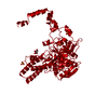
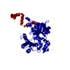
- PDB-6u0p: Crystal structure of PieE, the flavin-dependent monooxygenase inv... -
+ Open data

- Basic information
| Entry | Database: PDB / ID: 6u0p | ||||||
|---|---|---|---|---|---|---|---|
| Title | Crystal structure of PieE, the flavin-dependent monooxygenase involved in the biosynthesis of piericidin A1 | ||||||
 Components Components | 2,4-dichlorophenol 6-monooxygenase | ||||||
 Keywords Keywords | FLAVOPROTEIN / flavin-dependent monooxygenase | ||||||
| Function / homology |  Function and homology information Function and homology informationoxidoreductase activity, acting on paired donors, with incorporation or reduction of molecular oxygen, NAD(P)H as one donor, and incorporation of one atom of oxygen / FAD binding Similarity search - Function | ||||||
| Biological species |  Streptomyces sp. SCSIO 03032 (bacteria) Streptomyces sp. SCSIO 03032 (bacteria) | ||||||
| Method |  X-RAY DIFFRACTION / SYNCHROTRON / MOLECULAR REPLACEMENT / Resolution: 2.02 Å X-RAY DIFFRACTION / SYNCHROTRON / MOLECULAR REPLACEMENT / Resolution: 2.02 Å | ||||||
 Authors Authors | Shi, R. / Manenda, M. / Picard, M.-E. | ||||||
| Funding support |  Canada, 1items Canada, 1items
| ||||||
 Citation Citation | Journal: J.Biol.Chem. / Year: 2020 Title: Structural analyses of the Group A flavin-dependent monooxygenase PieE reveal a sliding FAD cofactor conformation bridging OUT and IN conformations. Authors: Manenda, M.S. / Picard, M.E. / Zhang, L. / Cyr, N. / Zhu, X. / Barma, J. / Pascal, J.M. / Couture, M. / Zhang, C. / Shi, R. | ||||||
| History |
|
- Structure visualization
| Structure viewer | Molecule: MolmilJmol/JSmol |
|---|
- Downloads & links
-Download
| PDBx/mmCIF format | 6u0p.cif.gz | 729.8 KB | Display | PDBx/mmCIF format |
|---|---|---|---|---|
| PDB format | pdb6u0p.ent.gz | 595.5 KB | Display | PDB format |
| PDBx/mmJSON format | 6u0p.json.gz | Tree view | PDBx/mmJSON format | |
| Others |  Other downloads Other downloads |
-Validation report
| Arichive directory | https://data.pdbj.org/pub/pdb/validation_reports/u0/6u0pftp://data.pdbj.org/pub/pdb/validation_reports/u0/6u0p | HTTPS FTP |
|---|
-Related structure data
| Related structure data | 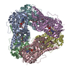 6u0sC 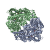 4z2uS S: Starting model for refinement C: citing same article ( |
|---|---|
| Similar structure data |


-Links
 PDBj
PDBj- Assembly
| Deposited unit | 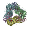
| ||||||||
|---|---|---|---|---|---|---|---|---|---|
| 1 |
| ||||||||
| Unit cell |
|
-Components
-Protein , 1 types, 6 molecules ABCDEF
| #1: Protein | Mass: 64754.434 Da / Num. of mol.: 6 Source method: isolated from a genetically manipulated source Source: (gene. exp.) Streptomyces sp. SCSIO 03032 (bacteria)Gene: CAG99_26330 / Plasmid: pET28a / Production host: |
|---|
-Non-polymers , 6 types, 2393 molecules 


| #2: Chemical | ChemComp-FAD /  Mass: 785.550 Da / Num. of mol.: 6 / Source method: obtained synthetically / Formula: C27H33N9O15P2 / Feature type: SUBJECT OF INVESTIGATION / Comment: FAD*YM Mass: 785.550 Da / Num. of mol.: 6 / Source method: obtained synthetically / Formula: C27H33N9O15P2 / Feature type: SUBJECT OF INVESTIGATION / Comment: FAD*YM#3: Chemical | ChemComp-PGE /  Mass: 150.173 Da / Num. of mol.: 12 / Source method: obtained synthetically / Formula: C6H14O4 Mass: 150.173 Da / Num. of mol.: 12 / Source method: obtained synthetically / Formula: C6H14O4#4: Chemical | ChemComp-1PE /  Mass: 238.278 Da / Num. of mol.: 6 / Source method: obtained synthetically / Formula: C10H22O6 / Comment: precipitant*YM Mass: 238.278 Da / Num. of mol.: 6 / Source method: obtained synthetically / Formula: C10H22O6 / Comment: precipitant*YM#5: Chemical | ChemComp-CL /  Mass: 35.453 Da / Num. of mol.: 10 / Source method: obtained synthetically / Formula: Cl Mass: 35.453 Da / Num. of mol.: 10 / Source method: obtained synthetically / Formula: Cl#6: Chemical | ChemComp-GOL /  Mass: 92.094 Da / Num. of mol.: 17 / Source method: obtained synthetically / Formula: C3H8O3 Mass: 92.094 Da / Num. of mol.: 17 / Source method: obtained synthetically / Formula: C3H8O3#7: Water | ChemComp-HOH / | Mass: 18.015 Da / Num. of mol.: 2342 / Source method: isolated from a natural source / Formula: H2O |
|---|
-Details
| Has ligand of interest | Y |
|---|
-Experimental details
-Experiment
| Experiment | Method: X-RAY DIFFRACTION / Number of used crystals: 1 |
|---|
- Sample preparation
| Crystal | Density Matthews: 2.68 Å3/Da / Density % sol: 54.03 % / Mosaicity: 0.37 ° |
|---|---|
| Crystal grow | Temperature: 295 K / Method: microbatch / pH: 7.5 / Details: 30% PEG 400, 0.2M MgCl2, and 0.1 HEPES pH 7.5 |
-Data collection
| Diffraction | Mean temperature: 100 K / Serial crystal experiment: N | ||||||||||||||||||||||||||||||
|---|---|---|---|---|---|---|---|---|---|---|---|---|---|---|---|---|---|---|---|---|---|---|---|---|---|---|---|---|---|---|---|
| Diffraction source | Source: SYNCHROTRON / Site: CLSI / Beamline: 08ID-1 / Wavelength: 0.9795 Å | ||||||||||||||||||||||||||||||
| Detector | Type: MAR CCD 130 mm / Detector: CCD / Date: Jan 8, 2016 | ||||||||||||||||||||||||||||||
| Radiation | Protocol: SINGLE WAVELENGTH / Monochromatic (M) / Laue (L): M / Scattering type: x-ray | ||||||||||||||||||||||||||||||
| Radiation wavelength | Wavelength: 0.9795 Å / Relative weight: 1 | ||||||||||||||||||||||||||||||
| Reflection | Resolution: 2.02→49.34 Å / Num. obs: 270573 / % possible obs: 99.8 % / Redundancy: 4.7 % / CC1/2: 0.994 / Rmerge(I) obs: 0.109 / Rpim(I) all: 0.054 / Rrim(I) all: 0.122 / Net I/σ(I): 8.8 / Num. measured all: 1275602 / Scaling rejects: 559 | ||||||||||||||||||||||||||||||
| Reflection shell | Diffraction-ID: 1
|
- Processing
| Software |
| ||||||||||||||||||||||||||||||||||||||||||||||||||||||||||||
|---|---|---|---|---|---|---|---|---|---|---|---|---|---|---|---|---|---|---|---|---|---|---|---|---|---|---|---|---|---|---|---|---|---|---|---|---|---|---|---|---|---|---|---|---|---|---|---|---|---|---|---|---|---|---|---|---|---|---|---|---|---|
| Refinement | Method to determine structure: MOLECULAR REPLACEMENT Starting model: 4Z2U Resolution: 2.02→49.34 Å / Cor.coef. Fo:Fc: 0.966 / Cor.coef. Fo:Fc free: 0.952 / SU B: 4.039 / SU ML: 0.107 / SU R Cruickshank DPI: 0.1613 / Cross valid method: THROUGHOUT / σ(F): 0 / ESU R: 0.161 / ESU R Free: 0.142 Details: HYDROGENS HAVE BEEN ADDED IN THE RIDING POSITIONS U VALUES : REFINED INDIVIDUALLY
| ||||||||||||||||||||||||||||||||||||||||||||||||||||||||||||
| Solvent computation | Ion probe radii: 0.8 Å / Shrinkage radii: 0.8 Å / VDW probe radii: 1.2 Å | ||||||||||||||||||||||||||||||||||||||||||||||||||||||||||||
| Displacement parameters | Biso max: 123.52 Å2 / Biso mean: 34.364 Å2 / Biso min: 14.03 Å2
| ||||||||||||||||||||||||||||||||||||||||||||||||||||||||||||
| Refinement step | Cycle: final / Resolution: 2.02→49.34 Å
| ||||||||||||||||||||||||||||||||||||||||||||||||||||||||||||
| Refine LS restraints |
| ||||||||||||||||||||||||||||||||||||||||||||||||||||||||||||
| LS refinement shell | Resolution: 2.02→2.072 Å / Rfactor Rfree error: 0 / Total num. of bins used: 20
|
