 Movie
Movie Controller

[English] 日本語
 Yorodumi
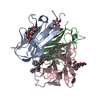
- PDB-6tum: Crystal structure of glutathione s-transferase PA1623 from Pseudo... -
+ Open data

- Basic information
| Entry | Database: PDB / ID: 6tum | ||||||
|---|---|---|---|---|---|---|---|
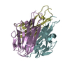
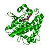
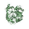
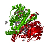
| Title | Crystal structure of glutathione s-transferase PA1623 from Pseudomonas aeruginosa PACS2 complexed with tartrate | ||||||
 Components Components | Glutathione S-transferase | ||||||
 Keywords Keywords | TRANSFERASE / Glutathione-S-Transferase / Detoxification / Oxidoreductase / Nu-class | ||||||
| Function / homology |  Function and homology information Function and homology information | ||||||
| Biological species |   Pseudomonas aeruginosa (bacteria) Pseudomonas aeruginosa (bacteria) | ||||||
| Method |  X-RAY DIFFRACTION / SYNCHROTRON / MOLECULAR REPLACEMENT / Resolution: 1.48 Å X-RAY DIFFRACTION / SYNCHROTRON / MOLECULAR REPLACEMENT / Resolution: 1.48 Å | ||||||
 Authors Authors | Feiler, C.G. / Blankenfeldt, W. | ||||||
 Citation Citation | Journal: To Be Published Title: Crystal structure of glutathione s-transferase PA1623 from Pseudomonas aeruginosa PACS2 complexed with tartrate Authors: Feiler, C.G. / Blankenfeldt, W. | ||||||
| History |
|
- Structure visualization
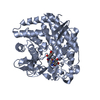
| Structure viewer | Molecule: MolmilJmol/JSmol |
|---|
- Downloads & links
-Download
| PDBx/mmCIF format | 6tum.cif.gz | 261.2 KB | Display | PDBx/mmCIF format |
|---|---|---|---|---|
| PDB format | pdb6tum.ent.gz | 217.2 KB | Display | PDB format |
| PDBx/mmJSON format | 6tum.json.gz | Tree view | PDBx/mmJSON format | |
| Others |  Other downloads Other downloads |
-Validation report
| Arichive directory | https://data.pdbj.org/pub/pdb/validation_reports/tu/6tumftp://data.pdbj.org/pub/pdb/validation_reports/tu/6tum | HTTPS FTP |
|---|
-Related structure data
| Related structure data |  4eciS S: Starting model for refinement |
|---|---|
| Similar structure data |


-Links
 PDBj
PDBj


- Assembly
| Deposited unit | 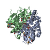
| ||||||||
|---|---|---|---|---|---|---|---|---|---|
| 1 |
| ||||||||
| Unit cell |
| ||||||||
| Components on special symmetry positions |
|
-Components
| #1: Protein | Mass: 25339.738 Da / Num. of mol.: 2 Source method: isolated from a genetically manipulated source Source: (gene. exp.) Pseudomonas aeruginosa (bacteria) / Gene: yfcG_4, NCTC13628_04787 / Production host: #2: Chemical | ChemComp-TAR / |   Mass: 150.087 Da / Num. of mol.: 1 / Source method: obtained synthetically / Formula: C4H6O6 Mass: 150.087 Da / Num. of mol.: 1 / Source method: obtained synthetically / Formula: C4H6O6#3: Chemical | ChemComp-TLA / |   Mass: 150.087 Da / Num. of mol.: 1 / Source method: obtained synthetically / Formula: C4H6O6 Mass: 150.087 Da / Num. of mol.: 1 / Source method: obtained synthetically / Formula: C4H6O6#4: Water | ChemComp-HOH / |  Mass: 18.015 Da / Num. of mol.: 420 / Source method: isolated from a natural source / Formula: H2O Mass: 18.015 Da / Num. of mol.: 420 / Source method: isolated from a natural source / Formula: H2OHas ligand of interest | N | Has protein modification | Y | |
|---|
-Experimental details
-Experiment
| Experiment | Method: X-RAY DIFFRACTION / Number of used crystals: 1 |
|---|
- Sample preparation
| Crystal | Density Matthews: 2.31 Å3/Da / Density % sol: 46.74 % |
|---|---|
| Crystal grow | Temperature: 293 K / Method: vapor diffusion, hanging drop / pH: 6.5 Details: 0.1 M BisTris pH 6.5, 0.1 M sodium potassium tartrate, 22 % PEG 3350 PH range: 6-7 |
-Data collection
| Diffraction | Mean temperature: 100 K / Serial crystal experiment: N |
|---|---|
| Diffraction source | Source: SYNCHROTRON / Site: BESSY  / Beamline: 14.1 / Wavelength: 0.91841 Å / Beamline: 14.1 / Wavelength: 0.91841 Å |
| Detector | Type: MARMOSAIC 225 mm CCD / Detector: CCD / Date: May 30, 2012 |
| Radiation | Monochromator: Si111 / Protocol: SINGLE WAVELENGTH / Monochromatic (M) / Laue (L): M / Scattering type: x-ray |
| Radiation wavelength | Wavelength: 0.91841 Å / Relative weight: 1 |
| Reflection | Resolution: 1.48→43.62 Å / Num. obs: 72280 / % possible obs: 99.47 % / Redundancy: 4.1 % / Biso Wilson estimate: 21.48 Å2 / CC1/2: 0.999 / Rmerge(I) obs: 0.077 / Rpim(I) all: 0.043 / Rrim(I) all: 0.088 / Net I/σ(I): 13.86 |
| Reflection shell | Resolution: 1.48→1.533 Å / Redundancy: 3.9 % / Rmerge(I) obs: 1.49 / Mean I/σ(I) obs: 0.99 / Num. unique obs: 7043 / CC1/2: 0.5 / Rpim(I) all: 0.86 / Rrim(I) all: 1.72 / % possible all: 98.15 |
- Processing
| Software |
| ||||||||||||||||||||||||||||||||||||||||||||||||||||||||||||||||||||||||||||||||||||||||||||||||||||||||||||||||
|---|---|---|---|---|---|---|---|---|---|---|---|---|---|---|---|---|---|---|---|---|---|---|---|---|---|---|---|---|---|---|---|---|---|---|---|---|---|---|---|---|---|---|---|---|---|---|---|---|---|---|---|---|---|---|---|---|---|---|---|---|---|---|---|---|---|---|---|---|---|---|---|---|---|---|---|---|---|---|---|---|---|---|---|---|---|---|---|---|---|---|---|---|---|---|---|---|---|---|---|---|---|---|---|---|---|---|---|---|---|---|---|---|---|
| Refinement | Method to determine structure: MOLECULAR REPLACEMENT Starting model: 4ECI Resolution: 1.48→43.62 Å / SU ML: 0.21 / Cross valid method: THROUGHOUT / σ(F): 1.35 / Phase error: 24.51
| ||||||||||||||||||||||||||||||||||||||||||||||||||||||||||||||||||||||||||||||||||||||||||||||||||||||||||||||||
| Solvent computation | Shrinkage radii: 0.9 Å / VDW probe radii: 1.11 Å | ||||||||||||||||||||||||||||||||||||||||||||||||||||||||||||||||||||||||||||||||||||||||||||||||||||||||||||||||
| Displacement parameters | Biso max: 119.42 Å2 / Biso mean: 25.7114 Å2 / Biso min: 10.89 Å2 | ||||||||||||||||||||||||||||||||||||||||||||||||||||||||||||||||||||||||||||||||||||||||||||||||||||||||||||||||
| Refinement step | Cycle: final / Resolution: 1.48→43.62 Å
| ||||||||||||||||||||||||||||||||||||||||||||||||||||||||||||||||||||||||||||||||||||||||||||||||||||||||||||||||
| LS refinement shell | Refine-ID: X-RAY DIFFRACTION / Rfactor Rfree error: 0 / Total num. of bins used: 15
|
