 Movie
Movie Controller


+ Open data

- Basic information
| Entry | Database: PDB / ID: 6tjc | |||||||||||||||
|---|---|---|---|---|---|---|---|---|---|---|---|---|---|---|---|---|
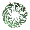
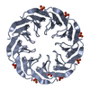
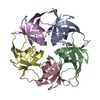
| Title | Crystal structure of the computationally designed Cake3 protein | |||||||||||||||
 Components Components | Cake3 | |||||||||||||||
 Keywords Keywords | DE NOVO PROTEIN / Beta-propeller / computationally designed / symmetrical / repeat protein | |||||||||||||||
| Function / homology | PHOSPHATE ION Function and homology information Function and homology information | |||||||||||||||
| Biological species | synthetic construct (others) | |||||||||||||||
| Method |  X-RAY DIFFRACTION / SYNCHROTRON / MOLECULAR REPLACEMENT / Resolution: 1.9 Å X-RAY DIFFRACTION / SYNCHROTRON / MOLECULAR REPLACEMENT / Resolution: 1.9 Å | |||||||||||||||
 Authors Authors | Laier, I. / Mylemans, B. / Voet, A.R.D. / Noguchi, H. | |||||||||||||||
| Funding support |  Belgium, 4items Belgium, 4items
| |||||||||||||||
 Citation Citation | Journal: Febs J. / Year: 2021 Title: Structural plasticity of a designer protein sheds light on beta-propeller protein evolution. Authors: Mylemans, B. / Laier, I. / Kamata, K. / Akashi, S. / Noguchi, H. / Tame, J.R.H. / Voet, A.R.D. | |||||||||||||||
| History |
|
- Structure visualization
| Structure viewer | Molecule: MolmilJmol/JSmol |
|---|
- Downloads & links
-Download
| PDBx/mmCIF format | 6tjc.cif.gz | 94.1 KB | Display | PDBx/mmCIF format |
|---|---|---|---|---|
| PDB format | pdb6tjc.ent.gz | 65.1 KB | Display | PDB format |
| PDBx/mmJSON format | 6tjc.json.gz | Tree view | PDBx/mmJSON format | |
| Others |  Other downloads Other downloads |
-Validation report
| Arichive directory | https://data.pdbj.org/pub/pdb/validation_reports/tj/6tjcftp://data.pdbj.org/pub/pdb/validation_reports/tj/6tjc | HTTPS FTP |
|---|
-Related structure data
| Related structure data |  6tjbC  6tjdC  6tjeC 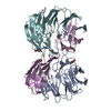 6tjfC 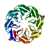 6tjgC  6tjhC 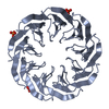 6tjiC C: citing same article ( |
|---|---|
| Similar structure data |


-Links
 PDBj
PDBj
- Assembly
| Deposited unit | 
| ||||||||||||
|---|---|---|---|---|---|---|---|---|---|---|---|---|---|
| 1 |
| ||||||||||||
| Unit cell |
|
-Components
| #1: Protein | Mass: 13315.628 Da / Num. of mol.: 3 Source method: isolated from a genetically manipulated source Source: (gene. exp.) synthetic construct (others) / Production host:  #2: Chemical | ChemComp-PO4 /   Mass: 94.971 Da / Num. of mol.: 4 / Source method: obtained synthetically / Formula: PO4 Mass: 94.971 Da / Num. of mol.: 4 / Source method: obtained synthetically / Formula: PO4#3: Chemical | ChemComp-GOL /   Mass: 92.094 Da / Num. of mol.: 4 / Source method: isolated from a natural source / Formula: C3H8O3 Mass: 92.094 Da / Num. of mol.: 4 / Source method: isolated from a natural source / Formula: C3H8O3#4: Water | ChemComp-HOH / |  Mass: 18.015 Da / Num. of mol.: 240 / Source method: isolated from a natural source / Formula: H2O Mass: 18.015 Da / Num. of mol.: 240 / Source method: isolated from a natural source / Formula: H2OHas ligand of interest | N | |
|---|
-Experimental details
-Experiment
| Experiment | Method: X-RAY DIFFRACTION / Number of used crystals: 1 |
|---|
- Sample preparation
| Crystal | Density Matthews: 2.27 Å3/Da / Density % sol: 45.85 % |
|---|---|
| Crystal grow | Temperature: 293.15 K / Method: vapor diffusion, sitting drop / Details: 0.05M Potassium phosphate, 20% PEG8000 |
-Data collection
| Diffraction | Mean temperature: 100 K / Serial crystal experiment: N |
|---|---|
| Diffraction source | Source: SYNCHROTRON / Site: Diamond  / Beamline: I03 / Wavelength: 0.9795 Å / Beamline: I03 / Wavelength: 0.9795 Å |
| Detector | Type: DECTRIS PILATUS3 6M / Detector: PIXEL / Date: Nov 26, 2018 |
| Radiation | Protocol: SINGLE WAVELENGTH / Monochromatic (M) / Laue (L): M / Scattering type: x-ray |
| Radiation wavelength | Wavelength: 0.9795 Å / Relative weight: 1 |
| Reflection | Resolution: 1.9→46.36 Å / Num. obs: 28542 / % possible obs: 100 % / Redundancy: 8.6 % / Biso Wilson estimate: 28 Å2 / CC1/2: 0.999 / Rmerge(I) obs: 0.055 / Rpim(I) all: 0.02 / Rrim(I) all: 0.059 / Χ2: 0.95 / Net I/σ(I): 21.8 |
| Reflection shell | Resolution: 1.9→1.94 Å / Redundancy: 5.6 % / Rmerge(I) obs: 0.387 / Mean I/σ(I) obs: 5.6 / Num. unique obs: 1793 / CC1/2: 0.957 / Rpim(I) all: 0.132 / Rrim(I) all: 0.409 / Χ2: 0.78 / % possible all: 100 |
- Processing
| Software |
| |||||||||||||||||||||||||||||||||||||||||||||||||||||||||||||||||||||||||||||
|---|---|---|---|---|---|---|---|---|---|---|---|---|---|---|---|---|---|---|---|---|---|---|---|---|---|---|---|---|---|---|---|---|---|---|---|---|---|---|---|---|---|---|---|---|---|---|---|---|---|---|---|---|---|---|---|---|---|---|---|---|---|---|---|---|---|---|---|---|---|---|---|---|---|---|---|---|---|---|
| Refinement | Method to determine structure: MOLECULAR REPLACEMENT Starting model: Computational design Resolution: 1.9→46.36 Å / SU ML: 0.1529 / Cross valid method: FREE R-VALUE / σ(F): 1.36 / Phase error: 19.6721 Stereochemistry target values: GeoStd + Monomer Library + CDL v1.2
| |||||||||||||||||||||||||||||||||||||||||||||||||||||||||||||||||||||||||||||
| Solvent computation | Shrinkage radii: 0.9 Å / VDW probe radii: 1.11 Å / Solvent model: FLAT BULK SOLVENT MODEL | |||||||||||||||||||||||||||||||||||||||||||||||||||||||||||||||||||||||||||||
| Displacement parameters | Biso mean: 28.83 Å2 | |||||||||||||||||||||||||||||||||||||||||||||||||||||||||||||||||||||||||||||
| Refinement step | Cycle: LAST / Resolution: 1.9→46.36 Å
| |||||||||||||||||||||||||||||||||||||||||||||||||||||||||||||||||||||||||||||
| Refine LS restraints |
| |||||||||||||||||||||||||||||||||||||||||||||||||||||||||||||||||||||||||||||
| LS refinement shell |
|
