 Movie
Movie Controller

[English] 日本語
 Yorodumi
- PDB-6tb8: Dye Type Peroxidase Aa from Streptomyces lividans: spectroscopica... -
+ Open data

- Basic information
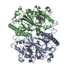
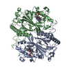
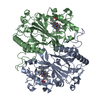
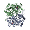
| Entry | Database: PDB / ID: 6tb8 | ||||||
|---|---|---|---|---|---|---|---|
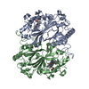
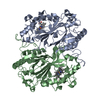
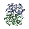
| Title | Dye Type Peroxidase Aa from Streptomyces lividans: spectroscopically-validated ferric state | ||||||
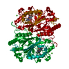
 Components Components | Deferrochelatase/peroxidase | ||||||
 Keywords Keywords | OXIDOREDUCTASE / peroxidase / metalloprotein / iron / dye type | ||||||
| Function / homology |  Function and homology information Function and homology informationiron import into cell / protoporphyrin ferrochelatase activity / cell envelope / Oxidoreductases; Acting on a peroxide as acceptor; Peroxidases / peroxidase activity / heme binding / metal ion binding / cytosol Similarity search - Function | ||||||
| Biological species |  Streptomyces lividans 1326 (bacteria) Streptomyces lividans 1326 (bacteria) | ||||||
| Method |  X-RAY DIFFRACTION / SYNCHROTRON / MOLECULAR REPLACEMENT / Resolution: 1.8 Å X-RAY DIFFRACTION / SYNCHROTRON / MOLECULAR REPLACEMENT / Resolution: 1.8 Å | ||||||
 Authors Authors | Lucic, M. / Moreno-Chicano, T.M. / Hough, M.A. / Dworkowski, F.S.N. / Worrall, J.A.R. | ||||||
 Citation Citation | Journal: Dalton Trans / Year: 2020 Title: A subtle structural change in the distal haem pocket has a remarkable effect on tuning hydrogen peroxide reactivity in dye decolourising peroxidases from Streptomyces lividans. Authors: Lucic, M. / Chaplin, A.K. / Moreno-Chicano, T. / Dworkowski, F.S.N. / Wilson, M.T. / Svistunenko, D.A. / Hough, M.A. / Worrall, J.A.R. | ||||||
| History |
|
- Structure visualization
| Structure viewer | Molecule: MolmilJmol/JSmol |
|---|
- Downloads & links
-Download
| PDBx/mmCIF format | 6tb8.cif.gz | 173.4 KB | Display | PDBx/mmCIF format |
|---|---|---|---|---|
| PDB format | pdb6tb8.ent.gz | 132.7 KB | Display | PDB format |
| PDBx/mmJSON format | 6tb8.json.gz | Tree view | PDBx/mmJSON format | |
| Others |  Other downloads Other downloads |
-Validation report
| Arichive directory | https://data.pdbj.org/pub/pdb/validation_reports/tb/6tb8ftp://data.pdbj.org/pub/pdb/validation_reports/tb/6tb8 | HTTPS FTP |
|---|
-Related structure data
| Related structure data | 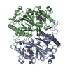 6i43S S: Starting model for refinement |
|---|---|
| Similar structure data |


-Links
 PDBj
PDBj

- Assembly
| Deposited unit | 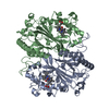
| ||||||||
|---|---|---|---|---|---|---|---|---|---|
| 1 |
| ||||||||
| Unit cell |
|
-Components
| #1: Protein | Mass: 40256.020 Da / Num. of mol.: 2 Source method: isolated from a genetically manipulated source Source: (gene. exp.) Streptomyces lividans 1326 (bacteria) / Gene: SLIV_26340 / Plasmid: pet28a / Production host: References: UniProt: A0A076MF68, UniProt: Q9RKQ2*PLUS, Oxidoreductases; Acting on a peroxide as acceptor; Peroxidases #2: Chemical |   Mass: 616.487 Da / Num. of mol.: 2 / Source method: obtained synthetically / Formula: C34H32FeN4O4 Mass: 616.487 Da / Num. of mol.: 2 / Source method: obtained synthetically / Formula: C34H32FeN4O4#3: Water | ChemComp-HOH / |  Mass: 18.015 Da / Num. of mol.: 848 / Source method: isolated from a natural source / Formula: H2O Mass: 18.015 Da / Num. of mol.: 848 / Source method: isolated from a natural source / Formula: H2OHas ligand of interest | N | |
|---|
-Experimental details
-Experiment
| Experiment | Method: X-RAY DIFFRACTION / Number of used crystals: 1 |
|---|
- Sample preparation
| Crystal | Density Matthews: 2.11 Å3/Da / Density % sol: 41.59 % |
|---|---|
| Crystal grow | Temperature: 291 K / Method: batch mode / pH: 7 Details: 1:1 RATIO OF A 6.5 MG/ML DTPAA PROTEIN SOLUTION WITH A PRECIPITANT SOLUTION CONTAINING 20% PEG 6000, 100 MM HEPES PH 7.0, BATCH MODE, TEMPERATURE 291K |
-Data collection
| Diffraction | Mean temperature: 100 K / Serial crystal experiment: N |
|---|---|
| Diffraction source | Source: SYNCHROTRON / Site: SLS  / Beamline: X10SA / Wavelength: 0.8 Å / Beamline: X10SA / Wavelength: 0.8 Å |
| Detector | Type: DECTRIS PILATUS 6M-F / Detector: PIXEL / Date: Sep 28, 2018 |
| Radiation | Protocol: SINGLE WAVELENGTH / Monochromatic (M) / Laue (L): M / Scattering type: x-ray |
| Radiation wavelength | Wavelength: 0.8 Å / Relative weight: 1 |
| Reflection | Resolution: 1.8→48.71 Å / Num. obs: 61725 / % possible obs: 99.4 % / Redundancy: 4.5 % / CC1/2: 0.989 / Rmerge(I) obs: 0.176 / Rpim(I) all: 0.089 / Rrim(I) all: 0.198 / Net I/av σ(I): 6.3 / Net I/σ(I): 6.3 |
| Reflection shell | Resolution: 1.8→1.84 Å / Redundancy: 2.9 % / Rmerge(I) obs: 0.57 / Mean I/σ(I) obs: 1.6 / Num. unique obs: 3542 / CC1/2: 0.683 / Rpim(I) all: 0.365 / Rrim(I) all: 0.682 / % possible all: 96.1 |
- Processing
| Software |
| ||||||||||||||||||||||||||||||||||||||||||||||||||||||||||||||||||||||||||||||||||||||||||||||||||||||||||||||||||||||||||||||||||||
|---|---|---|---|---|---|---|---|---|---|---|---|---|---|---|---|---|---|---|---|---|---|---|---|---|---|---|---|---|---|---|---|---|---|---|---|---|---|---|---|---|---|---|---|---|---|---|---|---|---|---|---|---|---|---|---|---|---|---|---|---|---|---|---|---|---|---|---|---|---|---|---|---|---|---|---|---|---|---|---|---|---|---|---|---|---|---|---|---|---|---|---|---|---|---|---|---|---|---|---|---|---|---|---|---|---|---|---|---|---|---|---|---|---|---|---|---|---|---|---|---|---|---|---|---|---|---|---|---|---|---|---|---|---|
| Refinement | Method to determine structure: MOLECULAR REPLACEMENT Starting model: 6I43 Resolution: 1.8→43.679 Å / SU ML: 0.21 / Cross valid method: THROUGHOUT / σ(F): 1.34 / Phase error: 20.99
| ||||||||||||||||||||||||||||||||||||||||||||||||||||||||||||||||||||||||||||||||||||||||||||||||||||||||||||||||||||||||||||||||||||
| Solvent computation | Shrinkage radii: 0.9 Å / VDW probe radii: 1.11 Å | ||||||||||||||||||||||||||||||||||||||||||||||||||||||||||||||||||||||||||||||||||||||||||||||||||||||||||||||||||||||||||||||||||||
| Displacement parameters | Biso max: 73.53 Å2 / Biso mean: 20.281 Å2 / Biso min: 6.81 Å2 | ||||||||||||||||||||||||||||||||||||||||||||||||||||||||||||||||||||||||||||||||||||||||||||||||||||||||||||||||||||||||||||||||||||
| Refinement step | Cycle: final / Resolution: 1.8→43.679 Å
| ||||||||||||||||||||||||||||||||||||||||||||||||||||||||||||||||||||||||||||||||||||||||||||||||||||||||||||||||||||||||||||||||||||
| LS refinement shell | Refine-ID: X-RAY DIFFRACTION / Rfactor Rfree error: 0
|
