 Movie
Movie Controller


+ Open data

- Basic information
| Entry | Database: PDB / ID: 6sn9 | |||||||||||||||
|---|---|---|---|---|---|---|---|---|---|---|---|---|---|---|---|---|
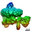
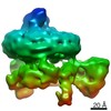
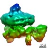
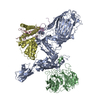
| Title | BamABCDE in MSP1D1 nanodisc ensemble 0-8 | |||||||||||||||
 Components Components |
| |||||||||||||||
 Keywords Keywords | MEMBRANE PROTEIN / Outer membrane / OMP / beta-barrel / folding / insertion | |||||||||||||||
| Function / homology |  Function and homology information Function and homology informationBam protein complex / Gram-negative-bacterium-type cell outer membrane assembly / protein insertion into membrane / cell outer membrane / protein-macromolecule adaptor activity / cell adhesion / response to antibiotic / cell surface / identical protein binding / membrane Similarity search - Function | |||||||||||||||
| Biological species |  | |||||||||||||||
| Method | ELECTRON MICROSCOPY / single particle reconstruction / cryo EM / Resolution: 9.8 Å | |||||||||||||||
 Authors Authors | Iadanza, M.G. / Ranson, N.A. / Radford, S.E. / Higgins, A.J. / Calabrese, A.N. / Schiffrin, B. / White, P. | |||||||||||||||
| Funding support |  United Kingdom, 4items United Kingdom, 4items
| |||||||||||||||
 Citation Citation | Journal: Commun Biol / Year: 2020 Title: Distortion of the bilayer and dynamics of the BAM complex in lipid nanodiscs. Authors: Matthew G Iadanza / Bob Schiffrin / Paul White / Matthew A Watson / Jim E Horne / Anna J Higgins / Antonio N Calabrese / David J Brockwell / Roman Tuma / Antreas C Kalli / Sheena E Radford / Neil A Ranson /  Abstract: The β-barrel assembly machinery (BAM) catalyses the folding and insertion of β-barrel outer membrane proteins (OMPs) into the outer membranes of Gram-negative bacteria by mechanisms that remain ...The β-barrel assembly machinery (BAM) catalyses the folding and insertion of β-barrel outer membrane proteins (OMPs) into the outer membranes of Gram-negative bacteria by mechanisms that remain unclear. Here, we present an ensemble of cryoEM structures of the E. coli BamABCDE (BAM) complex in lipid nanodiscs, determined using multi-body refinement techniques. These structures, supported by single-molecule FRET measurements, describe a range of motions in the BAM complex, mostly localised within the periplasmic region of the major subunit BamA. The β-barrel domain of BamA is in a 'lateral open' conformation in all of the determined structures, suggesting that this is the most energetically favourable species in this bilayer. Strikingly, the BAM-containing lipid nanodisc is deformed, especially around BAM's lateral gate. This distortion is also captured in molecular dynamics simulations, and provides direct structural evidence for the lipid 'disruptase' activity of BAM, suggested to be an important part of its functional mechanism. | |||||||||||||||
| History |
|
- Structure visualization
| Movie |
Movie viewer |
|---|---|
| Structure viewer | Molecule: MolmilJmol/JSmol |
- Downloads & links
-Download
| PDBx/mmCIF format | 6sn9.cif.gz | 274 KB | Display | PDBx/mmCIF format |
|---|---|---|---|---|
| PDB format | pdb6sn9.ent.gz | 217.1 KB | Display | PDB format |
| PDBx/mmJSON format | 6sn9.json.gz | Tree view | PDBx/mmJSON format | |
| Others |  Other downloads Other downloads |
-Validation report
| Summary document | 6sn9_validation.pdf.gz | 739.3 KB | Display | wwPDB validaton report |
|---|---|---|---|---|
| Full document | 6sn9_full_validation.pdf.gz | 780.3 KB | Display | |
| Data in XML | 6sn9_validation.xml.gz | 54.1 KB | Display | |
| Data in CIF | 6sn9_validation.cif.gz | 81.6 KB | Display | |
| Arichive directory | https://data.pdbj.org/pub/pdb/validation_reports/sn/6sn9ftp://data.pdbj.org/pub/pdb/validation_reports/sn/6sn9 | HTTPS FTP |
-Related structure data
| Related structure data |  10255MC  6smxC  6sn0C  6sn2C  6sn3C  6sn4C  6sn5C  6sn7C  6sn8C  6so7C  6so8C  6soaC  6sobC  6socC  6sogC  6sohC  6sojC C: citing same article ( M: map data used to model this data |
|---|---|
| Similar structure data |


-Links
 PDBj
PDBj


- Assembly
| Deposited unit | 
|
|---|---|
| 1 |
|
-Components
| #1: Protein | Mass: 87783.945 Da / Num. of mol.: 1 Source method: isolated from a genetically manipulated source Source: (gene. exp.) |
|---|---|
| #2: Protein | Mass: 39692.156 Da / Num. of mol.: 1 Source method: isolated from a genetically manipulated source Source: (gene. exp.) |
| #3: Protein | Mass: 6096.815 Da / Num. of mol.: 1 Source method: isolated from a genetically manipulated source Source: (gene. exp.) |
| #4: Protein | Mass: 25008.967 Da / Num. of mol.: 1 Source method: isolated from a genetically manipulated source Source: (gene. exp.) |
| #5: Protein | Mass: 9728.837 Da / Num. of mol.: 1 Source method: isolated from a genetically manipulated source Source: (gene. exp.) |
-Experimental details
-Experiment
| Experiment | Method: ELECTRON MICROSCOPY |
|---|---|
| EM experiment | Aggregation state: PARTICLE / 3D reconstruction method: single particle reconstruction |
- Sample preparation
| Component | Name: Bam complex / Type: COMPLEX Details: In MSP1D1 nanodisc with E. coli polar lipid extract Entity ID: all / Source: RECOMBINANT |
|---|---|
| Molecular weight | Value: 0.2 MDa / Experimental value: NO |
| Source (natural) | Organism: |
| Source (recombinant) | Organism: |
| Buffer solution | pH: 8 |
| Specimen | Embedding applied: NO / Shadowing applied: NO / Staining applied: NO / Vitrification applied: YES |
| Vitrification | Instrument: LEICA EM GP / Cryogen name: ETHANE / Humidity: 80 % / Chamber temperature: 281 K |
- Electron microscopy imaging
| Experimental equipment |  Model: Titan Krios / Image courtesy: FEI Company |
|---|---|
| Microscopy | Model: FEI TITAN KRIOS |
| Electron gun | Electron source:  FIELD EMISSION GUN / Accelerating voltage: 300 kV / Illumination mode: FLOOD BEAM FIELD EMISSION GUN / Accelerating voltage: 300 kV / Illumination mode: FLOOD BEAM |
| Electron lens | Mode: BRIGHT FIELD / Cs: 2.6 mm / Alignment procedure: COMA FREE |
| Specimen holder | Cryogen: NITROGEN / Specimen holder model: FEI TITAN KRIOS AUTOGRID HOLDER |
| Image recording | Average exposure time: 10 sec. / Electron dose: 40.75 e/Å2 / Detector mode: COUNTING / Film or detector model: GATAN K2 SUMMIT (4k x 4k) / Num. of grids imaged: 4 / Num. of real images: 15504 |
- Processing
| EM software |
| ||||||||||||||||||||||||||||||||||||||||
|---|---|---|---|---|---|---|---|---|---|---|---|---|---|---|---|---|---|---|---|---|---|---|---|---|---|---|---|---|---|---|---|---|---|---|---|---|---|---|---|---|---|
| CTF correction | Type: PHASE FLIPPING AND AMPLITUDE CORRECTION | ||||||||||||||||||||||||||||||||||||||||
| Symmetry | Point symmetry: C1 (asymmetric) | ||||||||||||||||||||||||||||||||||||||||
| 3D reconstruction | Resolution: 9.8 Å / Resolution method: FSC 0.143 CUT-OFF / Num. of particles: 20115 / Symmetry type: POINT | ||||||||||||||||||||||||||||||||||||||||
| Atomic model building | Protocol: FLEXIBLE FIT / Space: REAL | ||||||||||||||||||||||||||||||||||||||||
| Atomic model building | PDB-ID: 5LJO Accession code: 5LJO / Source name: PDB / Type: experimental model |

