 Movie
Movie Controller


+ Open data

- Basic information
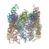
| Entry | Database: PDB / ID: 6sd0 | ||||||
|---|---|---|---|---|---|---|---|
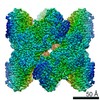
| Title | Structure of beta-galactosidase from Thermotoga maritima. | ||||||
 Components Components | Beta-galactosidase | ||||||
 Keywords Keywords | HYDROLASE / BETA-GALACTOSIDASE / GLYCOSIDE HYDROLASE / HYDROLYSIS / GALACTOOLIGOSACCHARIDE / THERMOTOGA MARITIMA / THERMORESISTANT / LACTOSE / GLYCOSYL HYDROLASE 2 FAMILY. SUGAR BINDING PROTEIN | ||||||
| Function / homology |  Function and homology information Function and homology informationlactose catabolic process / beta-galactosidase complex / beta-galactosidase / beta-galactosidase activity / carbohydrate binding Similarity search - Function | ||||||
| Biological species |   Thermotoga maritima MSB8 (bacteria) Thermotoga maritima MSB8 (bacteria) | ||||||
| Method |  X-RAY DIFFRACTION / SYNCHROTRON / MOLECULAR REPLACEMENT / Resolution: 3.7 Å X-RAY DIFFRACTION / SYNCHROTRON / MOLECULAR REPLACEMENT / Resolution: 3.7 Å | ||||||
 Authors Authors | Jimenez-Ortega, E. / Ramirez-Escudero, M. / Sanz-Aparicio, J. | ||||||
 Citation Citation | Journal: ACS Chem Biol / Year: 2020 Title: The cryo-EM Structure of β-Galactosidase: Quaternary Structure Guides Protein Engineering. Authors: Samuel Míguez Amil / Elena Jiménez-Ortega / Mercedes Ramírez-Escudero / David Talens-Perales / Julia Marín-Navarro / Julio Polaina / Julia Sanz-Aparicio / Rafael Fernandez-Leiro /  Abstract: Lactose intolerance is a common digestive disorder that affects a large proportion of the adult human population. The severity of the symptoms is highly variable, depending on the susceptibility to ...Lactose intolerance is a common digestive disorder that affects a large proportion of the adult human population. The severity of the symptoms is highly variable, depending on the susceptibility to the sugar and the amount digested. For that reason, enzymes that can be used for the production of lactose-free milk and milk derivatives have acquired singular biotechnological importance. One such case is β-galactosidase (TmLac). Here, we report the cryo-EM structure of TmLac at 2.0 Å resolution. The protein features a newly solved domain at its C-terminus, characteristic of the genus , which promotes a peculiar octameric arrangement. We have assessed the constraints imposed by the quaternary protein structure on the construction of hybrid versions of this GH2 enzyme. Carbohydrate binding modules (CBM) from the CBM2 and CBM9 families have been added at either the amino or carboxy terminus, and the structural and functional effects of such modifications have been analyzed. The results provide a basis for the rational design of hybrid enzymes that can be efficiently attached to different solid supports. | ||||||
| History |
|
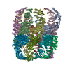
- Structure visualization
| Structure viewer | Molecule: MolmilJmol/JSmol |
|---|
- Downloads & links
-Download
| PDBx/mmCIF format | 6sd0.cif.gz | 892.1 KB | Display | PDBx/mmCIF format |
|---|---|---|---|---|
| PDB format | pdb6sd0.ent.gz | 731.1 KB | Display | PDB format |
| PDBx/mmJSON format | 6sd0.json.gz | Tree view | PDBx/mmJSON format | |
| Others |  Other downloads Other downloads |
-Validation report
| Summary document | 6sd0_validation.pdf.gz | 1 MB | Display | wwPDB validaton report |
|---|---|---|---|---|
| Full document | 6sd0_full_validation.pdf.gz | 1.1 MB | Display | |
| Data in XML | 6sd0_validation.xml.gz | 141 KB | Display | |
| Data in CIF | 6sd0_validation.cif.gz | 188.6 KB | Display | |
| Arichive directory | https://data.pdbj.org/pub/pdb/validation_reports/sd/6sd0ftp://data.pdbj.org/pub/pdb/validation_reports/sd/6sd0 | HTTPS FTP |
-Related structure data
| Related structure data |  6s6zC 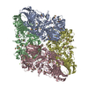 1dp0S S: Starting model for refinement C: citing same article ( |
|---|---|
| Similar structure data |


-Links
 PDBj
PDBj


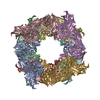
- Assembly
| Deposited unit | 
| |||||||||||||||||||||||||||||||||||||||||||||||||||||||||||||||||
|---|---|---|---|---|---|---|---|---|---|---|---|---|---|---|---|---|---|---|---|---|---|---|---|---|---|---|---|---|---|---|---|---|---|---|---|---|---|---|---|---|---|---|---|---|---|---|---|---|---|---|---|---|---|---|---|---|---|---|---|---|---|---|---|---|---|---|
| 1 | 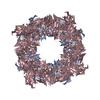
| |||||||||||||||||||||||||||||||||||||||||||||||||||||||||||||||||
| 2 | 
| |||||||||||||||||||||||||||||||||||||||||||||||||||||||||||||||||
| Unit cell |
| |||||||||||||||||||||||||||||||||||||||||||||||||||||||||||||||||
| Noncrystallographic symmetry (NCS) | NCS domain:
NCS domain segments:
NCS oper:
|
-Components
| #1: Protein | Mass: 127784.742 Da / Num. of mol.: 4 Source method: isolated from a genetically manipulated source Source: (gene. exp.) Thermotoga maritima MSB8 (bacteria) / Gene: lacZ, TM_1193 / Plasmid: pRARE2 / Production host: #2: Chemical | ChemComp-MG /   Mass: 24.305 Da / Num. of mol.: 4 / Source method: obtained synthetically / Formula: Mg / Feature type: SUBJECT OF INVESTIGATION Mass: 24.305 Da / Num. of mol.: 4 / Source method: obtained synthetically / Formula: Mg / Feature type: SUBJECT OF INVESTIGATION#3: Water | ChemComp-HOH / |  Mass: 18.015 Da / Num. of mol.: 3 / Source method: isolated from a natural source / Formula: H2O Mass: 18.015 Da / Num. of mol.: 3 / Source method: isolated from a natural source / Formula: H2OHas ligand of interest | Y | |
|---|
-Experimental details
-Experiment
| Experiment | Method: X-RAY DIFFRACTION / Number of used crystals: 1 |
|---|
- Sample preparation
| Crystal | Density Matthews: 3.59 Å3/Da / Density % sol: 65.7 % / Description: Crystal bar |
|---|---|
| Crystal grow | Temperature: 291 K / Method: vapor diffusion, sitting drop / pH: 7.5 Details: 12% PEG 3350, 0.1 M Bis-Tris propane pH 8.5, 0.2 M NaNO3, 2% MPD, 8 mM n-Nonyl-b-D-thiomaltoside. The crystals were enhanced by the microseeding technique. |
-Data collection
| Diffraction | Mean temperature: 100 K / Serial crystal experiment: N | |||||||||||||||
|---|---|---|---|---|---|---|---|---|---|---|---|---|---|---|---|---|
| Diffraction source | Source: SYNCHROTRON / Site: ALBA / Beamline: XALOC / Wavelength: 0.97947 Å | |||||||||||||||
| Detector | Type: DECTRIS PILATUS 6M / Detector: PIXEL / Date: Jun 4, 2014 / Details: KB focusing mirrors | |||||||||||||||
| Radiation | Monochromator: Si(111) channel-cut, cryocooled / Protocol: SINGLE WAVELENGTH / Monochromatic (M) / Laue (L): M / Scattering type: x-ray | |||||||||||||||
| Radiation wavelength | Wavelength: 0.97947 Å / Relative weight: 1 | |||||||||||||||
| Reflection twin |
| |||||||||||||||
| Reflection | Resolution: 3.7→40 Å / Num. all: 442617 / Num. obs: 76050 / % possible obs: 99.8 % / Redundancy: 5.8 % / CC1/2: 0.996 / Rmerge(I) obs: 0.165 / Rpim(I) all: 0.075 / Rrim(I) all: 0.182 / Net I/σ(I): 10.1 | |||||||||||||||
| Reflection shell | Resolution: 3.7→3.78 Å / Redundancy: 6 % / Rmerge(I) obs: 1.74 / Num. unique obs: 26943 / CC1/2: 0.581 / Rpim(I) all: 0.771 / Rrim(I) all: 1.906 / % possible all: 99.9 |
- Processing
| Software |
| ||||||||||||||||||||||||||||||||||||||||||||||||||||||||||||||||||||||||||||||
|---|---|---|---|---|---|---|---|---|---|---|---|---|---|---|---|---|---|---|---|---|---|---|---|---|---|---|---|---|---|---|---|---|---|---|---|---|---|---|---|---|---|---|---|---|---|---|---|---|---|---|---|---|---|---|---|---|---|---|---|---|---|---|---|---|---|---|---|---|---|---|---|---|---|---|---|---|---|---|---|
| Refinement | Method to determine structure: MOLECULAR REPLACEMENT Starting model: 1DP0 Resolution: 3.7→40 Å / Cor.coef. Fo:Fc: 0.924 / Cor.coef. Fo:Fc free: 0.889 / SU B: 41.163 / SU ML: 0.623 / Cross valid method: THROUGHOUT / σ(F): 0 / ESU R Free: 0.161 Details: HYDROGENS HAVE BEEN ADDED IN THE RIDING POSITIONS U VALUES : REFINED INDIVIDUALLY
| ||||||||||||||||||||||||||||||||||||||||||||||||||||||||||||||||||||||||||||||
| Solvent computation | Ion probe radii: 0.8 Å / Shrinkage radii: 0.8 Å / VDW probe radii: 1.2 Å | ||||||||||||||||||||||||||||||||||||||||||||||||||||||||||||||||||||||||||||||
| Displacement parameters | Biso max: 276.79 Å2 / Biso mean: 146.58 Å2 / Biso min: 38.45 Å2
| ||||||||||||||||||||||||||||||||||||||||||||||||||||||||||||||||||||||||||||||
| Refinement step | Cycle: final / Resolution: 3.7→40 Å
| ||||||||||||||||||||||||||||||||||||||||||||||||||||||||||||||||||||||||||||||
| Refine LS restraints |
| ||||||||||||||||||||||||||||||||||||||||||||||||||||||||||||||||||||||||||||||
| Refine LS restraints NCS | Ens-ID: 1 / Refine-ID: X-RAY DIFFRACTION
| ||||||||||||||||||||||||||||||||||||||||||||||||||||||||||||||||||||||||||||||
| LS refinement shell | Resolution: 3.7→3.796 Å / Rfactor Rfree error: 0 / Total num. of bins used: 20
|
