 Movie
Movie Controller

[English] 日本語
 Yorodumi
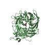
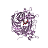
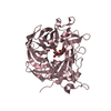
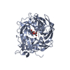
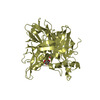
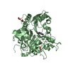
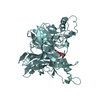
- PDB-6rv5: X-ray structure of the levansucrase from Erwinia tasmaniensis in ... -
+ Open data

- Basic information
| Entry | Database: PDB / ID: 6rv5 | |||||||||
|---|---|---|---|---|---|---|---|---|---|---|
| Title | X-ray structure of the levansucrase from Erwinia tasmaniensis in complex with levanbiose | |||||||||
 Components Components | Levansucrase (Beta-D-fructofuranosyl transferase) | |||||||||
 Keywords Keywords | TRANSFERASE / Levansucrase / levanbiose / Fructosyltransferase / Fructans production. | |||||||||
| Function / homology |  Function and homology information Function and homology informationlevansucrase / levansucrase activity / carbohydrate utilization / metal ion binding Similarity search - Function | |||||||||
| Biological species |  Erwinia tasmaniensis (bacteria) Erwinia tasmaniensis (bacteria) | |||||||||
| Method |  X-RAY DIFFRACTION / SYNCHROTRON / MOLECULAR REPLACEMENT / Resolution: 1.58 Å X-RAY DIFFRACTION / SYNCHROTRON / MOLECULAR REPLACEMENT / Resolution: 1.58 Å | |||||||||
 Authors Authors | Polsinelli, I. / Caliandro, R. / Demitri, N. / Benini, S. | |||||||||
 Citation Citation | Journal: Int J Mol Sci / Year: 2019 Title: The Structure of Sucrose-Soaked Levansucrase Crystals fromErwinia tasmaniensisreveals a Binding Pocket for Levanbiose. Authors: Polsinelli, I. / Caliandro, R. / Demitri, N. / Benini, S. #1: Journal: Int.J.Biol.Macromol. / Year: 2019Title: Comparison of the Levansucrase from the epiphyte Erwinia tasmaniensis vs its homologue from the phytopathogen Erwinia amylovora. Authors: Polsinelli, I. / Caliandro, R. / Salomone-Stagni, M. / Demitri, N. / Rejzek, M. / Field, R.A. / Benini, S. | |||||||||
| History |
|
- Structure visualization
| Structure viewer | Molecule: MolmilJmol/JSmol |
|---|
- Downloads & links
-Download
| PDBx/mmCIF format | 6rv5.cif.gz | 207.5 KB | Display | PDBx/mmCIF format |
|---|---|---|---|---|
| PDB format | pdb6rv5.ent.gz | 162.6 KB | Display | PDB format |
| PDBx/mmJSON format | 6rv5.json.gz | Tree view | PDBx/mmJSON format | |
| Others |  Other downloads Other downloads |
-Validation report
| Summary document | 6rv5_validation.pdf.gz | 914.2 KB | Display | wwPDB validaton report |
|---|---|---|---|---|
| Full document | 6rv5_full_validation.pdf.gz | 917.6 KB | Display | |
| Data in XML | 6rv5_validation.xml.gz | 22.1 KB | Display | |
| Data in CIF | 6rv5_validation.cif.gz | 33.7 KB | Display | |
| Arichive directory | https://data.pdbj.org/pub/pdb/validation_reports/rv/6rv5ftp://data.pdbj.org/pub/pdb/validation_reports/rv/6rv5 | HTTPS FTP |
-Related structure data
| Related structure data | 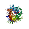 6frwS S: Starting model for refinement |
|---|---|
| Similar structure data |


-Links
 PDBj
PDBj- Assembly
| Deposited unit | 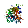
| ||||||||
|---|---|---|---|---|---|---|---|---|---|
| 1 |
| ||||||||
| Unit cell |
|
-Components
| #1: Protein | Mass: 46139.234 Da / Num. of mol.: 1 Source method: isolated from a genetically manipulated source Source: (gene. exp.) Erwinia tasmaniensis (strain DSM 17950 / CIP 109463 / Et1/99) (bacteria)Strain: DSM 17950 / CIP 109463 / Et1/99 / Gene: lsc, ETA_34670 / Production host: | ||||
|---|---|---|---|---|---|
| #2: Polysaccharide | beta-D-fructofuranose-(2-6)-beta-D-fructofuranose / levanbiose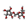  Source method: isolated from a genetically manipulated source Details: oligosaccharide / References: levanbiose | ||||
| #3: Chemical | ChemComp-GOL /   Mass: 92.094 Da / Num. of mol.: 11 / Source method: obtained synthetically / Formula: C3H8O3 Mass: 92.094 Da / Num. of mol.: 11 / Source method: obtained synthetically / Formula: C3H8O3#4: Chemical |   Mass: 65.409 Da / Num. of mol.: 2 / Source method: obtained synthetically / Formula: Zn Mass: 65.409 Da / Num. of mol.: 2 / Source method: obtained synthetically / Formula: Zn#5: Water | ChemComp-HOH / |  Mass: 18.015 Da / Num. of mol.: 407 / Source method: isolated from a natural source / Formula: H2O Mass: 18.015 Da / Num. of mol.: 407 / Source method: isolated from a natural source / Formula: H2O |
-Experimental details
-Experiment
| Experiment | Method: X-RAY DIFFRACTION / Number of used crystals: 1 |
|---|
- Sample preparation
| Crystal | Density Matthews: 2.58 Å3/Da / Density % sol: 52.36 % |
|---|---|
| Crystal grow | Temperature: 293.15 K / Method: vapor diffusion, hanging drop / pH: 7.5 Details: 28% Glycerol, 14% PEG 4000, 1.5mM Manganese(II) chloride tetrahydrate, 1.5 mM Cobalt(II) chloride hexahydrate , 1.5 mM Nickel(II) chloride hexahydrate, 1.5 mM Zinc acetate dihydrate crystals ...Details: 28% Glycerol, 14% PEG 4000, 1.5mM Manganese(II) chloride tetrahydrate, 1.5 mM Cobalt(II) chloride hexahydrate , 1.5 mM Nickel(II) chloride hexahydrate, 1.5 mM Zinc acetate dihydrate crystals soaked in Sucrose 500 mM |
-Data collection
| Diffraction | Mean temperature: 100 K / Serial crystal experiment: N |
|---|---|
| Diffraction source | Source: SYNCHROTRON / Site: ELETTRA  / Beamline: 5.2R / Wavelength: 1 Å / Beamline: 5.2R / Wavelength: 1 Å |
| Detector | Type: DECTRIS PILATUS 2M / Detector: PIXEL / Date: Dec 13, 2017 |
| Radiation | Protocol: SINGLE WAVELENGTH / Monochromatic (M) / Laue (L): M / Scattering type: x-ray |
| Radiation wavelength | Wavelength: 1 Å / Relative weight: 1 |
| Reflection | Resolution: 1.45→90.42 Å / Num. obs: 66162 / % possible obs: 99.47 % / Redundancy: 1.3 % / CC1/2: 0.999 / Rmerge(I) obs: 0.02668 / Rpim(I) all: 0.02668 / Rrim(I) all: 0.03773 / Net I/σ(I): 2.04 |
| Reflection shell | Resolution: 1.58→1.64 Å / Rmerge(I) obs: 0.2867 / Num. unique obs: 6517 / CC1/2: 0.888 / Rpim(I) all: 0.2867 / Rrim(I) all: 0.4055 |
- Processing
| Software |
| ||||||||||||||||||||
|---|---|---|---|---|---|---|---|---|---|---|---|---|---|---|---|---|---|---|---|---|---|
| Refinement | Method to determine structure: MOLECULAR REPLACEMENT Starting model: 6FRW Resolution: 1.58→45.26 Å / Cor.coef. Fo:Fc: 0.98 / Cor.coef. Fo:Fc free: 0.966 / Cross valid method: THROUGHOUT / Details: HYDROGENS HAVE BEEN ADDED IN THE RIDING POSITIONS
| ||||||||||||||||||||
| Displacement parameters | Biso mean: 23.661 Å2 | ||||||||||||||||||||
| Refinement step | Cycle: 1 / Resolution: 1.58→45.26 Å
| ||||||||||||||||||||
| LS refinement shell | Resolution: 1.58→1.621 Å
|
