 Movie
Movie Controller

[English] 日本語
 Yorodumi
- PDB-6rs4: Structure of tabersonine synthase - an alpha-beta hydrolase from ... -
+ Open data

- Basic information
| Entry | Database: PDB / ID: 6rs4 | |||||||||
|---|---|---|---|---|---|---|---|---|---|---|
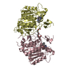
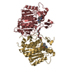
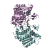
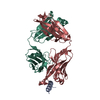
| Title | Structure of tabersonine synthase - an alpha-beta hydrolase from Catharanthus roseus | |||||||||
 Components Components | Tabersonine synthase | |||||||||
 Keywords Keywords | HYDROLASE / alkaloid / TABERSONINE / natural product / biosynthesis / alpha/beta hydrolase fold | |||||||||
| Function / homology |  Function and homology information Function and homology informationLyases / alkaloid metabolic process / lyase activity / hydrolase activity / nucleus / cytosol Similarity search - Function | |||||||||
| Biological species |  Catharanthus roseus (Madagascar periwinkle) Catharanthus roseus (Madagascar periwinkle) | |||||||||
| Method |  X-RAY DIFFRACTION / SYNCHROTRON / MOLECULAR REPLACEMENT / Resolution: 1.3 Å X-RAY DIFFRACTION / SYNCHROTRON / MOLECULAR REPLACEMENT / Resolution: 1.3 Å | |||||||||
 Authors Authors | Caputi, L. / Franke, J. / Bussey, K. / Farrow, S.C. / Curcino Vieira, I.J. / Stevenson, C.E.M. / Lawson, D.M. / O'Connor, S.E. | |||||||||
| Funding support |  Belgium, Belgium,  United Kingdom, 2items United Kingdom, 2items
| |||||||||
 Citation Citation | Journal: Nat.Chem.Biol. / Year: 2020 Title: Structural basis of cycloaddition in biosynthesis of iboga and aspidosperma alkaloids. Authors: Caputi, L. / Franke, J. / Bussey, K. / Farrow, S.C. / Vieira, I.J.C. / Stevenson, C.E.M. / Lawson, D.M. / O'Connor, S.E. | |||||||||
| History |
|
- Structure visualization
| Structure viewer | Molecule: MolmilJmol/JSmol |
|---|
- Downloads & links
-Download
| PDBx/mmCIF format | 6rs4.cif.gz | 299.1 KB | Display | PDBx/mmCIF format |
|---|---|---|---|---|
| PDB format | pdb6rs4.ent.gz | 242.1 KB | Display | PDB format |
| PDBx/mmJSON format | 6rs4.json.gz | Tree view | PDBx/mmJSON format | |
| Others |  Other downloads Other downloads |
-Validation report
| Arichive directory | https://data.pdbj.org/pub/pdb/validation_reports/rs/6rs4ftp://data.pdbj.org/pub/pdb/validation_reports/rs/6rs4 | HTTPS FTP |
|---|
-Related structure data
| Related structure data | 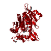 6rj8C 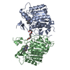 6rt8C  2o7rS S: Starting model for refinement C: citing same article ( |
|---|---|
| Similar structure data |


-Links
 PDBj
PDBj- Assembly
| Deposited unit | 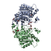
| ||||||||
|---|---|---|---|---|---|---|---|---|---|
| 1 |
| ||||||||
| Unit cell |
|
-Components
| #1: Protein | Mass: 36256.051 Da / Num. of mol.: 2 Mutation: Native N-terminal MET is replaced by a GLY-PRO dipeptide left over from cleavage of the affinity tag Source method: isolated from a genetically manipulated source Source: (gene. exp.) Catharanthus roseus (Madagascar periwinkle)Gene: TS, HL2 / Plasmid: pOPINF / Production host:  #2: Chemical | ChemComp-EDO /   Mass: 62.068 Da / Num. of mol.: 11 / Source method: obtained synthetically / Formula: C2H6O2 Mass: 62.068 Da / Num. of mol.: 11 / Source method: obtained synthetically / Formula: C2H6O2#3: Water | ChemComp-HOH / |  Mass: 18.015 Da / Num. of mol.: 699 / Source method: isolated from a natural source / Formula: H2O Mass: 18.015 Da / Num. of mol.: 699 / Source method: isolated from a natural source / Formula: H2O |
|---|
-Experimental details
-Experiment
| Experiment | Method: X-RAY DIFFRACTION / Number of used crystals: 1 |
|---|
- Sample preparation
| Crystal | Density Matthews: 2.54 Å3/Da / Density % sol: 51.66 % |
|---|---|
| Crystal grow | Temperature: 293 K / Method: vapor diffusion, sitting drop / pH: 5 / Details: NULL / PH range: NULL / Temp details: NULL |
-Data collection
| Diffraction | Mean temperature: 100 K / Serial crystal experiment: N | ||||||||||||||||||||||||||||||
|---|---|---|---|---|---|---|---|---|---|---|---|---|---|---|---|---|---|---|---|---|---|---|---|---|---|---|---|---|---|---|---|
| Diffraction source | Source: SYNCHROTRON / Site: Diamond / Beamline: I03 / Wavelength: 0.9795 Å | ||||||||||||||||||||||||||||||
| Detector | Type: DECTRIS PILATUS3 6M / Detector: PIXEL / Date: Jul 17, 2016 | ||||||||||||||||||||||||||||||
| Radiation | Protocol: SINGLE WAVELENGTH / Monochromatic (M) / Laue (L): M / Scattering type: x-ray | ||||||||||||||||||||||||||||||
| Radiation wavelength | Wavelength: 0.9795 Å / Relative weight: 1 | ||||||||||||||||||||||||||||||
| Reflection | Resolution: 1.3→59.95 Å / Num. obs: 177427 / % possible obs: 100 % / Redundancy: 6.7 % / Biso Wilson estimate: 10.6 Å2 / CC1/2: 0.998 / Rmerge(I) obs: 0.111 / Rpim(I) all: 0.046 / Rrim(I) all: 0.12 / Net I/σ(I): 10.8 | ||||||||||||||||||||||||||||||
| Reflection shell | Diffraction-ID: 1
|
- Processing
| Software |
| |||||||||||||||||||||||||||||||||||||||||||||||||||||||||||||||||
|---|---|---|---|---|---|---|---|---|---|---|---|---|---|---|---|---|---|---|---|---|---|---|---|---|---|---|---|---|---|---|---|---|---|---|---|---|---|---|---|---|---|---|---|---|---|---|---|---|---|---|---|---|---|---|---|---|---|---|---|---|---|---|---|---|---|---|
| Refinement | Method to determine structure: MOLECULAR REPLACEMENT Starting model: 2O7R Resolution: 1.3→59.95 Å / Cor.coef. Fo:Fc: 0.981 / Cor.coef. Fo:Fc free: 0.975 / SU B: 1.907 / SU ML: 0.033 / SU R Cruickshank DPI: 0.0402 / Cross valid method: THROUGHOUT / σ(F): 0 / ESU R: 0.04 / ESU R Free: 0.04 Details: HYDROGENS HAVE BEEN ADDED IN THE RIDING POSITIONS U VALUES : REFINED INDIVIDUALLY
| |||||||||||||||||||||||||||||||||||||||||||||||||||||||||||||||||
| Solvent computation | Ion probe radii: 0.8 Å / Shrinkage radii: 0.8 Å / VDW probe radii: 1.2 Å | |||||||||||||||||||||||||||||||||||||||||||||||||||||||||||||||||
| Displacement parameters | Biso max: 77.48 Å2 / Biso mean: 16.166 Å2 / Biso min: 6.81 Å2
| |||||||||||||||||||||||||||||||||||||||||||||||||||||||||||||||||
| Refinement step | Cycle: final / Resolution: 1.3→59.95 Å
| |||||||||||||||||||||||||||||||||||||||||||||||||||||||||||||||||
| Refine LS restraints |
| |||||||||||||||||||||||||||||||||||||||||||||||||||||||||||||||||
| LS refinement shell | Resolution: 1.3→1.334 Å / Rfactor Rfree error: 0 / Total num. of bins used: 20
|
