 Movie
Movie Controller


+ Open data

- Basic information
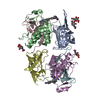
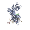
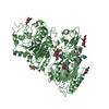
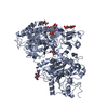
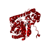
| Entry | Database: PDB / ID: 6rn6 | ||||||
|---|---|---|---|---|---|---|---|
| Title | DPP1 in complex with inhibitor | ||||||
 Components Components | (Dipeptidyl peptidase ...) x 3 | ||||||
 Keywords Keywords | HYDROLASE / DIPEPTIDYL PEPTIDASE I / INHIBITOR COMPLEX / Cathepsin C | ||||||
| Function / homology |  Function and homology information Function and homology informationdipeptidyl-peptidase I / peptidase activator activity involved in apoptotic process / : / Cargo concentration in the ER / COPII-coated ER to Golgi transport vesicle / dipeptidyl-peptidase activity / COPII-mediated vesicle transport / chloride ion binding / phosphatase binding / endoplasmic reticulum-Golgi intermediate compartment membrane ...dipeptidyl-peptidase I / peptidase activator activity involved in apoptotic process / : / Cargo concentration in the ER / COPII-coated ER to Golgi transport vesicle / dipeptidyl-peptidase activity / COPII-mediated vesicle transport / chloride ion binding / phosphatase binding / endoplasmic reticulum-Golgi intermediate compartment membrane / cysteine-type peptidase activity / MHC class II antigen presentation / : / T cell mediated cytotoxicity / azurophil granule lumen / protein-folding chaperone binding / extracellular matrix / lysosome / immune response / endoplasmic reticulum lumen / serine-type endopeptidase activity / cysteine-type endopeptidase activity / Neutrophil degranulation / proteolysis / : / extracellular exosome / extracellular region / membrane / identical protein binding Similarity search - Function | ||||||
| Biological species |  Homo sapiens (human) Homo sapiens (human) | ||||||
| Method |  X-RAY DIFFRACTION / SYNCHROTRON / MOLECULAR REPLACEMENT / Resolution: 2.4 Å X-RAY DIFFRACTION / SYNCHROTRON / MOLECULAR REPLACEMENT / Resolution: 2.4 Å | ||||||
 Authors Authors | Kack, H. | ||||||
 Citation Citation | Journal: Acs Med.Chem.Lett. / Year: 2019 Title: DPP1 Inhibitors: Exploring the Role of Water in the S2 Pocket of DPP1 with Substituted Pyrrolidines. Authors: Kack, H. / Doyle, K. / Hughes, S.J. / Bodnarchuk, M.S. / Lonn, H. / Van De Poel, A. / Palmer, N. | ||||||
| History |
|
- Structure visualization
| Structure viewer | Molecule: MolmilJmol/JSmol |
|---|
- Downloads & links
-Download
| PDBx/mmCIF format | 6rn6.cif.gz | 291.3 KB | Display | PDBx/mmCIF format |
|---|---|---|---|---|
| PDB format | pdb6rn6.ent.gz | 237.2 KB | Display | PDB format |
| PDBx/mmJSON format | 6rn6.json.gz | Tree view | PDBx/mmJSON format | |
| Others |  Other downloads Other downloads |
-Validation report
| Arichive directory | https://data.pdbj.org/pub/pdb/validation_reports/rn/6rn6ftp://data.pdbj.org/pub/pdb/validation_reports/rn/6rn6 | HTTPS FTP |
|---|
-Related structure data
| Related structure data | 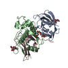 6rn7C 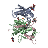 6rn9C 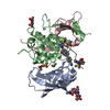 6rneC 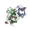 6rniC  1k3bS S: Starting model for refinement C: citing same article ( |
|---|---|
| Similar structure data |


-Links
 PDBj
PDBj


- Assembly
| Deposited unit | 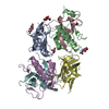
| ||||||||
|---|---|---|---|---|---|---|---|---|---|
| 1 |
| ||||||||
| Unit cell |
|
-Components
-Dipeptidyl peptidase ... , 3 types, 6 molecules ADBECF
| #1: Protein | Mass: 13500.163 Da / Num. of mol.: 2 Source method: isolated from a genetically manipulated source Source: (gene. exp.) Homo sapiens (human) / Gene: CTSC, CPPI / Production host:   Spodoptera frugiperda (fall armyworm) / References: UniProt: P53634, dipeptidyl-peptidase I Spodoptera frugiperda (fall armyworm) / References: UniProt: P53634, dipeptidyl-peptidase I#2: Protein | Mass: 18373.689 Da / Num. of mol.: 2 Source method: isolated from a genetically manipulated source Source: (gene. exp.) Homo sapiens (human) / Gene: CTSC, CPPI / Production host: Spodoptera frugiperda (fall armyworm) / References: UniProt: P53634, dipeptidyl-peptidase I#3: Protein | Mass: 7583.444 Da / Num. of mol.: 2 Source method: isolated from a genetically manipulated source Source: (gene. exp.) Homo sapiens (human) / Gene: CTSC, CPPI / Production host: Spodoptera frugiperda (fall armyworm) / References: UniProt: P53634, dipeptidyl-peptidase I |
|---|
-Sugars , 1 types, 3 molecules 
| #4: Sugar |  Type: D-saccharide, beta linking / Mass: 221.208 Da / Num. of mol.: 3 Type: D-saccharide, beta linking / Mass: 221.208 Da / Num. of mol.: 3Source method: isolated from a genetically manipulated source Formula: C8H15NO6 |
|---|
-Non-polymers , 4 types, 234 molecules 


| #5: Chemical | ChemComp-GOL /  Mass: 92.094 Da / Num. of mol.: 1 / Source method: obtained synthetically / Formula: C3H8O3 Mass: 92.094 Da / Num. of mol.: 1 / Source method: obtained synthetically / Formula: C3H8O3 | ||||
|---|---|---|---|---|---|
| #6: Chemical | ChemComp-CL /  Mass: 35.453 Da / Num. of mol.: 5 / Source method: obtained synthetically / Formula: Cl Mass: 35.453 Da / Num. of mol.: 5 / Source method: obtained synthetically / Formula: Cl#7: Chemical | ChemComp-K9Q / ( |  Mass: 339.431 Da / Num. of mol.: 1 / Source method: obtained synthetically / Formula: C20H25N3O2 Mass: 339.431 Da / Num. of mol.: 1 / Source method: obtained synthetically / Formula: C20H25N3O2#8: Water | ChemComp-HOH / | Mass: 18.015 Da / Num. of mol.: 227 / Source method: isolated from a natural source / Formula: H2O |
-Details
| Has protein modification | Y |
|---|
-Experimental details
-Experiment
| Experiment | Method: X-RAY DIFFRACTION / Number of used crystals: 1 |
|---|
- Sample preparation
| Crystal | Density Matthews: 2.98 Å3/Da / Density % sol: 58.7 % |
|---|---|
| Crystal grow | Temperature: 293 K / Method: vapor diffusion, hanging drop / pH: 5 Details: PEG 3350, 0.2 M Ammoniumsulphate, 0.1 M sodium acetate |
-Data collection
| Diffraction | Mean temperature: 100 K / Serial crystal experiment: N |
|---|---|
| Diffraction source | Source: SYNCHROTRON / Site: ESRF  / Beamline: ID23-2 / Wavelength: 0.873 Å / Beamline: ID23-2 / Wavelength: 0.873 Å |
| Detector | Type: ADSC QUANTUM 4 / Detector: CCD / Date: Jul 19, 2007 |
| Radiation | Protocol: SINGLE WAVELENGTH / Monochromatic (M) / Laue (L): M / Scattering type: x-ray |
| Radiation wavelength | Wavelength: 0.873 Å / Relative weight: 1 |
| Reflection | Resolution: 2.4→74.5 Å / Num. obs: 36889 / % possible obs: 99.5 % / Redundancy: 3.9 % / CC1/2: 0.996 / Rmerge(I) obs: 0.099 / Net I/σ(I): 11.3 |
| Reflection shell | Resolution: 2.4→2.42 Å / Redundancy: 3.9 % / Rmerge(I) obs: 0.763 / Mean I/σ(I) obs: 2.2 / Num. unique obs: 1786 / CC1/2: 0.704 / % possible all: 95.6 |
- Processing
| Software |
| |||||||||||||||||||||||||||||||||||||||||||||||||||||||||||||||||||||||||||||||||||||||||||||||||||||||||||||||||||||||||||||||||||||||||||||||||||||||||||||||||||||||||||||||
|---|---|---|---|---|---|---|---|---|---|---|---|---|---|---|---|---|---|---|---|---|---|---|---|---|---|---|---|---|---|---|---|---|---|---|---|---|---|---|---|---|---|---|---|---|---|---|---|---|---|---|---|---|---|---|---|---|---|---|---|---|---|---|---|---|---|---|---|---|---|---|---|---|---|---|---|---|---|---|---|---|---|---|---|---|---|---|---|---|---|---|---|---|---|---|---|---|---|---|---|---|---|---|---|---|---|---|---|---|---|---|---|---|---|---|---|---|---|---|---|---|---|---|---|---|---|---|---|---|---|---|---|---|---|---|---|---|---|---|---|---|---|---|---|---|---|---|---|---|---|---|---|---|---|---|---|---|---|---|---|---|---|---|---|---|---|---|---|---|---|---|---|---|---|---|---|---|
| Refinement | Method to determine structure: MOLECULAR REPLACEMENT Starting model: 1K3B Resolution: 2.4→14.87 Å / Cor.coef. Fo:Fc: 0.939 / Cor.coef. Fo:Fc free: 0.905 / SU R Cruickshank DPI: 0.272 / Cross valid method: THROUGHOUT / SU R Blow DPI: 0.282 / SU Rfree Blow DPI: 0.211 / SU Rfree Cruickshank DPI: 0.21
| |||||||||||||||||||||||||||||||||||||||||||||||||||||||||||||||||||||||||||||||||||||||||||||||||||||||||||||||||||||||||||||||||||||||||||||||||||||||||||||||||||||||||||||||
| Displacement parameters | Biso mean: 52.83 Å2
| |||||||||||||||||||||||||||||||||||||||||||||||||||||||||||||||||||||||||||||||||||||||||||||||||||||||||||||||||||||||||||||||||||||||||||||||||||||||||||||||||||||||||||||||
| Refine analyze | Luzzati coordinate error obs: 0.27 Å | |||||||||||||||||||||||||||||||||||||||||||||||||||||||||||||||||||||||||||||||||||||||||||||||||||||||||||||||||||||||||||||||||||||||||||||||||||||||||||||||||||||||||||||||
| Refinement step | Cycle: LAST / Resolution: 2.4→14.87 Å
| |||||||||||||||||||||||||||||||||||||||||||||||||||||||||||||||||||||||||||||||||||||||||||||||||||||||||||||||||||||||||||||||||||||||||||||||||||||||||||||||||||||||||||||||
| LS refinement shell | Highest resolution: 2.4 Å | |||||||||||||||||||||||||||||||||||||||||||||||||||||||||||||||||||||||||||||||||||||||||||||||||||||||||||||||||||||||||||||||||||||||||||||||||||||||||||||||||||||||||||||||
| Refinement TLS params. | Refine-ID: X-RAY DIFFRACTION
| |||||||||||||||||||||||||||||||||||||||||||||||||||||||||||||||||||||||||||||||||||||||||||||||||||||||||||||||||||||||||||||||||||||||||||||||||||||||||||||||||||||||||||||||
| Refinement TLS group |
|
