 Movie
Movie Controller

[English] 日本語
 Yorodumi
- PDB-6r8s: Crystal structure of aIF2gamma subunit I181K from archaeon Sulfol... -
+ Open data

- Basic information
| Entry | Database: PDB / ID: 6r8s | ||||||
|---|---|---|---|---|---|---|---|
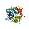
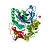
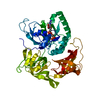
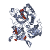
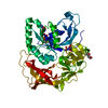
| Title | Crystal structure of aIF2gamma subunit I181K from archaeon Sulfolobus solfataricus complexed with GDPCP | ||||||
 Components Components | Translation initiation factor 2 subunit gamma | ||||||
 Keywords Keywords | TRANSLATION / INITIATION FACTOR 2 / GAMMA SUBUNIT / INITIATION OF THE TRANSLATION / NUCLEOTIDE BINDING / GDPCP / PROTEIN BIOSYNTHESIS | ||||||
| Function / homology |  Function and homology information Function and homology informationformation of translation preinitiation complex / protein-synthesizing GTPase / translation elongation factor activity / translation initiation factor activity / tRNA binding / GTPase activity / GTP binding / metal ion binding / cytosol Similarity search - Function | ||||||
| Biological species |   Saccharolobus solfataricus (archaea) Saccharolobus solfataricus (archaea) | ||||||
| Method |  X-RAY DIFFRACTION / SYNCHROTRON / MOLECULAR REPLACEMENT / Resolution: 2.18 Å X-RAY DIFFRACTION / SYNCHROTRON / MOLECULAR REPLACEMENT / Resolution: 2.18 Å | ||||||
 Authors Authors | Kravchenko, O. / Arkhipova, V. / Gabdulkhakov, A. / Stolboushkina, E. / Nikonov, O. / Garber, M. / Nikonov, S. | ||||||
 Citation Citation | Journal: To Be Published Title: Crystal structure of aIF2gamma subunit I181K from archaeon Sulfolobus solfataricus complexed with GDPCP Authors: Nikonov, O. / Kravchenko, O. / Stolboushkina, E. / Nevskaya, N. / Garber, M. / Nikonov, S. | ||||||
| History |
|
- Structure visualization
| Structure viewer | Molecule: MolmilJmol/JSmol |
|---|
- Downloads & links
-Download
| PDBx/mmCIF format | 6r8s.cif.gz | 111.9 KB | Display | PDBx/mmCIF format |
|---|---|---|---|---|
| PDB format | pdb6r8s.ent.gz | 82.8 KB | Display | PDB format |
| PDBx/mmJSON format | 6r8s.json.gz | Tree view | PDBx/mmJSON format | |
| Others |  Other downloads Other downloads |
-Validation report
| Arichive directory | https://data.pdbj.org/pub/pdb/validation_reports/r8/6r8sftp://data.pdbj.org/pub/pdb/validation_reports/r8/6r8s | HTTPS FTP |
|---|
-Related structure data
| Related structure data |  4nbsS S: Starting model for refinement |
|---|---|
| Similar structure data |


-Links
 PDBj
PDBj


- Assembly
| Deposited unit | 
| ||||||||
|---|---|---|---|---|---|---|---|---|---|
| 1 |
| ||||||||
| Unit cell |
|
-Components
| #1: Protein | Mass: 45865.250 Da / Num. of mol.: 1 / Mutation: I181K Source method: isolated from a genetically manipulated source Source: (gene. exp.) Saccharolobus solfataricus (archaea)Gene: eif2g, SSOP1_0394, SULA_1435, SULB_1436, SULC_1434, SULG_07135, SULH_07135, SULI_07135, SULM_07135, SULN_07135, SULO_07145, SULZ_07375 Production host:  | ||||
|---|---|---|---|---|---|
| #2: Chemical | ChemComp-GCP /   Mass: 521.208 Da / Num. of mol.: 1 / Source method: obtained synthetically / Formula: C11H18N5O13P3 / Comment: GMP-PCP, energy-carrying molecule analogue*YM Mass: 521.208 Da / Num. of mol.: 1 / Source method: obtained synthetically / Formula: C11H18N5O13P3 / Comment: GMP-PCP, energy-carrying molecule analogue*YM | ||||
| #3: Chemical | ChemComp-MG /   Mass: 24.305 Da / Num. of mol.: 1 / Source method: obtained synthetically / Formula: Mg Mass: 24.305 Da / Num. of mol.: 1 / Source method: obtained synthetically / Formula: Mg | ||||
| #4: Chemical | ChemComp-FMT /   Mass: 46.025 Da / Num. of mol.: 13 / Source method: obtained synthetically / Formula: CH2O2 Mass: 46.025 Da / Num. of mol.: 13 / Source method: obtained synthetically / Formula: CH2O2#5: Water | ChemComp-HOH / |  Mass: 18.015 Da / Num. of mol.: 479 / Source method: isolated from a natural source / Formula: H2O Mass: 18.015 Da / Num. of mol.: 479 / Source method: isolated from a natural source / Formula: H2OHas protein modification | Y | |
-Experimental details
-Experiment
| Experiment | Method: X-RAY DIFFRACTION / Number of used crystals: 1 |
|---|
- Sample preparation
| Crystal grow | Temperature: 301 K / Method: vapor diffusion, hanging drop Details: 17 mM TRIS-HCL pH7,5, 17 mM NaCl, 3,4mM 2-mercaptoethanol, 1,22 M sodium formate, 33,9 mM sodium cacodylate pH 6.5, 0,33% MMEPEG 5000, 1,7mM GDPCP |
|---|
-Data collection
| Diffraction | Mean temperature: 100 K / Serial crystal experiment: N |
|---|---|
| Diffraction source | Source: SYNCHROTRON / Site: BESSY  / Beamline: 14.1 / Wavelength: 0.918409 Å / Beamline: 14.1 / Wavelength: 0.918409 Å |
| Detector | Type: DECTRIS PILATUS3 6M / Detector: PIXEL / Date: Apr 13, 2014 |
| Radiation | Protocol: SINGLE WAVELENGTH / Monochromatic (M) / Laue (L): M / Scattering type: x-ray |
| Radiation wavelength | Wavelength: 0.918409 Å / Relative weight: 1 |
| Reflection | Resolution: 2.18→46.714 Å / Num. obs: 56432 / % possible obs: 100 % / Redundancy: 11.39 % / Net I/σ(I): 14.24 |
- Processing
| Software |
| ||||||||||||||||||||||||||||||||||||||||||||||||||||||||||||||||||||||||||||||||||||||||||||||||||||||||||||||||||||||||||||||||||||||||||||
|---|---|---|---|---|---|---|---|---|---|---|---|---|---|---|---|---|---|---|---|---|---|---|---|---|---|---|---|---|---|---|---|---|---|---|---|---|---|---|---|---|---|---|---|---|---|---|---|---|---|---|---|---|---|---|---|---|---|---|---|---|---|---|---|---|---|---|---|---|---|---|---|---|---|---|---|---|---|---|---|---|---|---|---|---|---|---|---|---|---|---|---|---|---|---|---|---|---|---|---|---|---|---|---|---|---|---|---|---|---|---|---|---|---|---|---|---|---|---|---|---|---|---|---|---|---|---|---|---|---|---|---|---|---|---|---|---|---|---|---|---|---|
| Refinement | Method to determine structure: MOLECULAR REPLACEMENT Starting model: 4NBS Resolution: 2.18→46.714 Å / SU ML: 0.2 / Cross valid method: FREE R-VALUE / σ(F): 1.35 / Phase error: 18.5
| ||||||||||||||||||||||||||||||||||||||||||||||||||||||||||||||||||||||||||||||||||||||||||||||||||||||||||||||||||||||||||||||||||||||||||||
| Solvent computation | Shrinkage radii: 0.9 Å / VDW probe radii: 1.11 Å | ||||||||||||||||||||||||||||||||||||||||||||||||||||||||||||||||||||||||||||||||||||||||||||||||||||||||||||||||||||||||||||||||||||||||||||
| Refinement step | Cycle: LAST / Resolution: 2.18→46.714 Å
| ||||||||||||||||||||||||||||||||||||||||||||||||||||||||||||||||||||||||||||||||||||||||||||||||||||||||||||||||||||||||||||||||||||||||||||
| Refine LS restraints |
| ||||||||||||||||||||||||||||||||||||||||||||||||||||||||||||||||||||||||||||||||||||||||||||||||||||||||||||||||||||||||||||||||||||||||||||
| LS refinement shell |
|
