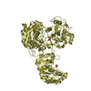
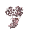
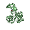
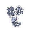
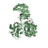
[English] 日本語
Yorodumi
- PDB-6qsn: Crystal structure of Yellow fever virus polymerase NS5A -

+
Open data


ID or keywords:

Loading...

-
Basic information

Entry
Database: PDB / ID: 6qsn
TitleCrystal structure of Yellow fever virus polymerase NS5A
ComponentsGenome polyprotein
KeywordsTRANSFERASE / virus / flavivirus / polymerase / RNA
Function / homology
Function and homology information


flavivirin / symbiont-mediated suppression of host JAK-STAT cascade via inhibition of STAT2 activity / double-stranded RNA binding / viral capsid / nucleoside-triphosphate phosphatase / mRNA (guanine-N7)-methyltransferase / methyltransferase cap1 / clathrin-dependent endocytosis of virus by host cell / mRNA (nucleoside-2'-O-)-methyltransferase activity / mRNA 5'-cap (guanine-N7-)-methyltransferase activity ...flavivirin / symbiont-mediated suppression of host JAK-STAT cascade via inhibition of STAT2 activity / double-stranded RNA binding / viral capsid / nucleoside-triphosphate phosphatase / mRNA (guanine-N7)-methyltransferase / methyltransferase cap1 / clathrin-dependent endocytosis of virus by host cell / mRNA (nucleoside-2'-O-)-methyltransferase activity / mRNA 5'-cap (guanine-N7-)-methyltransferase activity / RNA helicase activity / host cell endoplasmic reticulum membrane / host cell perinuclear region of cytoplasm / protein dimerization activity / symbiont-mediated suppression of host type I interferon-mediated signaling pathway / RNA helicase / induction by virus of host autophagy / RNA-directed RNA polymerase / viral RNA genome replication / serine-type endopeptidase activity / RNA-dependent RNA polymerase activity / virus-mediated perturbation of host defense response / fusion of virus membrane with host endosome membrane / viral envelope / host cell nucleus / virion attachment to host cell / GTP binding / structural molecule activity / virion membrane / ATP hydrolysis activity / proteolysis / extracellular region / ATP binding / membrane / metal ion binding
Similarity search - Function
: / Flavivirus capsid protein C superfamily / RNA-directed RNA polymerase, thumb domain, Flavivirus / Flavivirus RNA-directed RNA polymerase, thumb domain / Flavivirus non-structural protein NS2B / Flavivirus envelope glycoprotein E, stem/anchor domain / : / Flavivirus NS3 helicase, C-terminal helical domain / Genome polyprotein, Flavivirus / Flavivirus non-structural protein NS4A ...: / Flavivirus capsid protein C superfamily / RNA-directed RNA polymerase, thumb domain, Flavivirus / Flavivirus RNA-directed RNA polymerase, thumb domain / Flavivirus non-structural protein NS2B / Flavivirus envelope glycoprotein E, stem/anchor domain / : / Flavivirus NS3 helicase, C-terminal helical domain / Genome polyprotein, Flavivirus / Flavivirus non-structural protein NS4A / Flavivirus non-structural protein NS2B / Flavivirus capsid protein C / Flavivirus non-structural protein NS4B / mRNA cap 0/1 methyltransferase / Flavivirus capsid protein C / Flavivirus non-structural protein NS4B / Flavivirus non-structural protein NS4A / Flavivirus NS2B domain profile. / mRNA cap 0 and cap 1 methyltransferase (EC 2.1.1.56 and EC 2.1.1.57) domain profile. / Flavivirus non-structural protein NS2A / Flavivirus non-structural protein NS2A / Flavivirus NS3, petidase S7 / Peptidase S7, Flavivirus NS3 serine protease / Flavivirus NS3 protease (NS3pro) domain profile. / RNA-directed RNA polymerase, flavivirus / Flavivirus RNA-directed RNA polymerase, fingers and palm domains / Flavivirus non-structural Protein NS1 / Flavivirus non-structural protein NS1 / Envelope glycoprotein M, flavivirus / Flavivirus envelope glycoprotein M / Envelope glycoprotein M superfamily, flavivirus / Flavivirus polyprotein propeptide / Flavivirus polyprotein propeptide superfamily / Flavivirus polyprotein propeptide / Flaviviral glycoprotein E, central domain, subdomain 1 / Flaviviral glycoprotein E, central domain, subdomain 2 / Flavivirus envelope glycoprotein E, Stem/Anchor domain / Flavivirus glycoprotein E, immunoglobulin-like domain / Flavivirus envelope glycoprotein E, Stem/Anchor domain superfamily / Flavivirus glycoprotein, immunoglobulin-like domain / Flavivirus glycoprotein central and dimerisation domain / Flavivirus glycoprotein, central and dimerisation domains / Ribosomal RNA methyltransferase, FtsJ domain / FtsJ-like methyltransferase / Flavivirus/Alphavirus glycoprotein, immunoglobulin-like domain superfamily / Flavivirus glycoprotein, central and dimerisation domain superfamily / Flaviviral glycoprotein E, dimerisation domain / DEAD box, Flavivirus / Flavivirus DEAD domain / Vaccinia Virus protein VP39 / helicase superfamily c-terminal domain / Immunoglobulin E-set / Superfamilies 1 and 2 helicase C-terminal domain profile. / Superfamilies 1 and 2 helicase ATP-binding type-1 domain profile. / DEAD-like helicases superfamily / Helicase, C-terminal / Helicase superfamily 1/2, ATP-binding domain / RNA-directed RNA polymerase, catalytic domain / RdRp of positive ssRNA viruses catalytic domain profile. / S-adenosyl-L-methionine-dependent methyltransferase superfamily / Peptidase S1, PA clan / DNA/RNA polymerase superfamily / P-loop containing nucleoside triphosphate hydrolase / Rossmann fold / 3-Layer(aba) Sandwich / Alpha Beta
Similarity search - Domain/homology
S-ADENOSYL-L-HOMOCYSTEINE / Genome polyprotein
Similarity search - Component
Biological speciesYellow fever virus 17D
MethodX-RAY DIFFRACTION / SYNCHROTRON / Resolution: 3.004 Å
AuthorsBoura, E. / Dubankova, A.
Funding support Czech Republic, 1items
OrganizationGrant numberCountry
European Regional Development FundCZ.02.1.01/0.0/0.0/16_019/0000729 Czech Republic
CitationJournal: Antiviral Res. / Year: 2019
Title: Structure of the yellow fever NS5 protein reveals conserved drug targets shared among flaviviruses.
Authors: Dubankova, A. / Boura, E.
History
DepositionFeb 21, 2019Deposition site: PDBE / Processing site: PDBE
Revision 1.0Oct 16, 2019Provider: repository / Type: Initial release
Revision 1.1May 15, 2024Group: Data collection / Database references / Category: chem_comp_atom / chem_comp_bond / database_2
Item: _database_2.pdbx_DOI / _database_2.pdbx_database_accession

-
Structure visualization

Structure viewerMolecule:
MolmilJmol/JSmol

Downloads & links

-
Assembly

Deposited unit
A: Genome polyprotein
B: Genome polyprotein
hetero molecules


Theoretical massNumber of molelcules
Total (without water)209,90115
Polymers208,1982
Non-polymers1,70313
Water00
1
A: Genome polyprotein
hetero molecules


Theoretical massNumber of molelcules
Total (without water)104,9037
Polymers104,0991
Non-polymers8036
Water0
TypeNameSymmetry operationNumber
identity operation1_555x,y,z1
2
B: Genome polyprotein
hetero molecules


Theoretical massNumber of molelcules
Total (without water)104,9998
Polymers104,0991
Non-polymers8997
Water0
TypeNameSymmetry operationNumber
identity operation1_555x,y,z1
Unit cell
Length a, b, c (Å)108.285, 142.114, 152.177
Angle α, β, γ (deg.)90.00, 90.00, 90.00
Int Tables number19
Space group name H-MP212121

-
Components

#1: Protein Genome polyprotein


Mass: 104099.156 Da / Num. of mol.: 2
Source method: isolated from a genetically manipulated source
Source: (gene. exp.) Yellow fever virus 17D / Production host: Escherichia coli (E. coli)
References: UniProt: P03314, flavivirin, nucleoside-triphosphate phosphatase, RNA helicase, mRNA (guanine-N7)-methyltransferase, methyltransferase cap1, RNA-directed RNA polymerase
#2: Chemical ChemComp-SAH / S-ADENOSYL-L-HOMOCYSTEINE


Type: L-peptide linking / Mass: 384.411 Da / Num. of mol.: 2 / Source method: obtained synthetically / Formula: C14H20N6O5S
#3: Chemical
ChemComp-SO4 / SULFATE ION


Mass: 96.063 Da / Num. of mol.: 7 / Source method: obtained synthetically / Formula: SO4
#4: Chemical
ChemComp-ZN / ZINC ION


Mass: 65.409 Da / Num. of mol.: 4 / Source method: obtained synthetically / Formula: Zn

-
Experimental details

-
Experiment

ExperimentMethod: X-RAY DIFFRACTION / Number of used crystals: 1

-
Sample preparation

CrystalDensity Matthews: 2.81 Å3/Da / Density % sol: 56.26 %
Crystal growTemperature: 291 K / Method: vapor diffusion, sitting drop / pH: 8.5
Details: 80 mM Tris pH=8.5, 1.92M (NH4)2SO4, 20% (v/v) glycerol

-
Data collection

DiffractionMean temperature: 100 K / Serial crystal experiment: N
Diffraction sourceSource: SYNCHROTRON / Site: BESSY / Beamline: 14.2 / Wavelength: 0.9184 Å
DetectorType: DECTRIS PILATUS 2M / Detector: PIXEL / Date: Jan 18, 2019
RadiationProtocol: SINGLE WAVELENGTH / Monochromatic (M) / Laue (L): M / Scattering type: x-ray
Radiation wavelengthWavelength: 0.9184 Å / Relative weight: 1
ReflectionResolution: 3.004→48.011 Å / Num. obs: 47148 / % possible obs: 99.19 % / Redundancy: 8.3 % / CC1/2: 0.994 / Rpim(I) all: 0.081 / Net I/σ(I): 9.91
Reflection shellResolution: 3.004→3.11 Å / CC1/2: 0.419 / Rpim(I) all: 0.687

-
Processing

Software
NameVersionClassification
PHENIX(1.11.1_2575: ???)refinement
XDSdata reduction
XDSdata scaling
PHASERphasing
RefinementResolution: 3.004→48.011 Å / SU ML: 0.39 / Cross valid method: FREE R-VALUE / σ(F): 1.34 / Phase error: 26.35
RfactorNum. reflection% reflection
Rfree0.2706 2355 5 %
Rwork0.2207 --
obs0.2232 47121 99.23 %
Solvent computationShrinkage radii: 0.9 Å / VDW probe radii: 1.11 Å
Refinement stepCycle: LAST / Resolution: 3.004→48.011 Å
ProteinNucleic acidLigandSolventTotal
Num. atoms14227 0 91 0 14318
Refine LS restraints
Refine-IDTypeDev idealNumber
X-RAY DIFFRACTIONf_bond_d0.00214626
X-RAY DIFFRACTIONf_angle_d0.52419751
X-RAY DIFFRACTIONf_dihedral_angle_d8.5888757
X-RAY DIFFRACTIONf_chiral_restr0.042117
X-RAY DIFFRACTIONf_plane_restr0.0032511
LS refinement shell
Resolution (Å)Rfactor RfreeNum. reflection RfreeRfactor RworkNum. reflection RworkRefine-ID% reflection obs (%)
3.0044-3.06570.34741320.32932506X-RAY DIFFRACTION96
3.0657-3.13240.3371370.3082609X-RAY DIFFRACTION100
3.1324-3.20520.34731380.30322621X-RAY DIFFRACTION100
3.2052-3.28540.33221360.28712596X-RAY DIFFRACTION100
3.2854-3.37420.3131390.27992635X-RAY DIFFRACTION100
3.3742-3.47350.31341390.2662642X-RAY DIFFRACTION100
3.4735-3.58550.29121380.24582610X-RAY DIFFRACTION100
3.5855-3.71360.30641390.24342644X-RAY DIFFRACTION100
3.7136-3.86230.31681380.23712629X-RAY DIFFRACTION100
3.8623-4.0380.28871390.22652636X-RAY DIFFRACTION100
4.038-4.25070.26251330.20422519X-RAY DIFFRACTION95
4.2507-4.51690.25751380.19462639X-RAY DIFFRACTION100
4.5169-4.86530.21991400.18532660X-RAY DIFFRACTION100
4.8653-5.35430.2471410.20192670X-RAY DIFFRACTION100
5.3543-6.12780.29011420.2142693X-RAY DIFFRACTION100
6.1278-7.71520.24641430.2092719X-RAY DIFFRACTION100
7.7152-48.01710.2151430.17582738X-RAY DIFFRACTION96

+
About Yorodumi

-
News

-
Feb 9, 2022. New format data for meta-information of EMDB entries

New format data for meta-information of EMDB entries

  • Version 3 of the EMDB header file is now the official format.
  • The previous official version 1.9 will be removed from the archive.

Related info.:EMDB header

External links:wwPDB to switch to version 3 of the EMDB data model

-
Aug 12, 2020. Covid-19 info

Covid-19 info

URL: https://pdbjlvh1.pdbj.org/emnavi/covid19.php

New page: Covid-19 featured information page in EM Navigator.

Related info.:Covid-19 info / Mar 5, 2020. Novel coronavirus structure data

+
Mar 5, 2020. Novel coronavirus structure data

Novel coronavirus structure data

Related info.:Yorodumi Speices / Aug 12, 2020. Covid-19 info

External links:COVID-19 featured content - PDBj / Molecule of the Month (242):Coronavirus Proteases

+
Jan 31, 2019. EMDB accession codes are about to change! (news from PDBe EMDB page)

EMDB accession codes are about to change! (news from PDBe EMDB page)

  • The allocation of 4 digits for EMDB accession codes will soon come to an end. Whilst these codes will remain in use, new EMDB accession codes will include an additional digit and will expand incrementally as the available range of codes is exhausted. The current 4-digit format prefixed with “EMD-” (i.e. EMD-XXXX) will advance to a 5-digit format (i.e. EMD-XXXXX), and so on. It is currently estimated that the 4-digit codes will be depleted around Spring 2019, at which point the 5-digit format will come into force.
  • The EM Navigator/Yorodumi systems omit the EMD- prefix.

Related info.:Q: What is EMD? / ID/Accession-code notation in Yorodumi/EM Navigator

External links:EMDB Accession Codes are Changing Soon! / Contact to PDBj

+
Jul 12, 2017. Major update of PDB

Major update of PDB

  • wwPDB released updated PDB data conforming to the new PDBx/mmCIF dictionary.
  • This is a major update changing the version number from 4 to 5, and with Remediation, in which all the entries are updated.
  • In this update, many items about electron microscopy experimental information are reorganized (e.g. em_software).
  • Now, EM Navigator and Yorodumi are based on the updated data.

External links:wwPDB Remediation / Enriched Model Files Conforming to OneDep Data Standards Now Available in the PDB FTP Archive

-
Yorodumi

Thousand views of thousand structures

  • Yorodumi is a browser for structure data from EMDB, PDB, SASBDB, etc.
  • This page is also the successor to EM Navigator detail page, and also detail information page/front-end page for Omokage search.
  • The word "yorodu" (or yorozu) is an old Japanese word meaning "ten thousand". "mi" (miru) is to see.

Related info.:EMDB / PDB / SASBDB / Comparison of 3 databanks / Yorodumi Search / Aug 31, 2016. New EM Navigator & Yorodumi / Yorodumi Papers / Jmol/JSmol / Function and homology information / Changes in new EM Navigator and Yorodumi

Read more