 Movie
Movie Controller


+ Open data

- Basic information
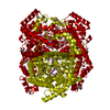
| Entry | Database: PDB / ID: 6qrr | ||||||
|---|---|---|---|---|---|---|---|
| Title | X-ray radiation dose series on xylose isomerase - 0.13 MGy | ||||||
 Components Components | Xylose isomerase | ||||||
 Keywords Keywords | METAL BINDING PROTEIN / Isomerase | ||||||
| Function / homology |  Function and homology information Function and homology informationxylose isomerase / D-xylose metabolic process / xylose isomerase activity / magnesium ion binding / identical protein binding / cytoplasm Similarity search - Function | ||||||
| Biological species |  Streptomyces rubiginosus (bacteria) Streptomyces rubiginosus (bacteria) | ||||||
| Method |  X-RAY DIFFRACTION / SYNCHROTRON / MOLECULAR REPLACEMENT / Resolution: 1.096 Å X-RAY DIFFRACTION / SYNCHROTRON / MOLECULAR REPLACEMENT / Resolution: 1.096 Å | ||||||
 Authors Authors | Taberman, H. / Bury, C.S. / van der Woerd, M.J. / Snell, E.H. / Garman, E.F. | ||||||
| Funding support |  Finland, 1items Finland, 1items
| ||||||
 Citation Citation | Journal: J.Synchrotron Radiat. / Year: 2019 Title: Structural knowledge or X-ray damage? A case study on xylose isomerase illustrating both. Authors: Taberman, H. / Bury, C.S. / van der Woerd, M.J. / Snell, E.H. / Garman, E.F. | ||||||
| History |
|
- Structure visualization
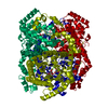
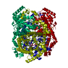
| Structure viewer | Molecule: MolmilJmol/JSmol |
|---|
- Downloads & links
-Download
| PDBx/mmCIF format | 6qrr.cif.gz | 219.3 KB | Display | PDBx/mmCIF format |
|---|---|---|---|---|
| PDB format | pdb6qrr.ent.gz | 173 KB | Display | PDB format |
| PDBx/mmJSON format | 6qrr.json.gz | Tree view | PDBx/mmJSON format | |
| Others |  Other downloads Other downloads |
-Validation report
| Summary document | 6qrr_validation.pdf.gz | 3.7 MB | Display | wwPDB validaton report |
|---|---|---|---|---|
| Full document | 6qrr_full_validation.pdf.gz | 3.7 MB | Display | |
| Data in XML | 6qrr_validation.xml.gz | 23.9 KB | Display | |
| Data in CIF | 6qrr_validation.cif.gz | 37.2 KB | Display | |
| Arichive directory | https://data.pdbj.org/pub/pdb/validation_reports/qr/6qrrftp://data.pdbj.org/pub/pdb/validation_reports/qr/6qrr | HTTPS FTP |
-Related structure data
| Related structure data |  6qrsC  6qrtC 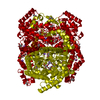 6qruC  6qrvC  6qrwC  6qrxC  6qryC  1muwS S: Starting model for refinement C: citing same article ( |
|---|---|
| Similar structure data |


-Links
 PDBj
PDBj

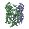
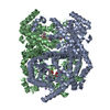
- Assembly
| Deposited unit | 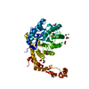
| ||||||||||||
|---|---|---|---|---|---|---|---|---|---|---|---|---|---|
| 1 | 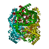
| ||||||||||||
| Unit cell |
| ||||||||||||
| Components on special symmetry positions |
|
-Components
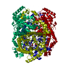
-Protein , 1 types, 1 molecules A
| #1: Protein | Mass: 43195.277 Da / Num. of mol.: 1 / Mutation: E186Q Source method: isolated from a genetically manipulated source Source: (gene. exp.) Streptomyces rubiginosus (bacteria) / Gene: xylA / Production host: |
|---|
-Non-polymers , 6 types, 545 molecules 


| #2: Chemical | ChemComp-EDO /  Mass: 62.068 Da / Num. of mol.: 12 / Source method: obtained synthetically / Formula: C2H6O2 Mass: 62.068 Da / Num. of mol.: 12 / Source method: obtained synthetically / Formula: C2H6O2#3: Chemical | ChemComp-IPA / |  Mass: 60.095 Da / Num. of mol.: 1 / Source method: obtained synthetically / Formula: C3H8O / Comment: alkaloid*YM Mass: 60.095 Da / Num. of mol.: 1 / Source method: obtained synthetically / Formula: C3H8O / Comment: alkaloid*YM#4: Chemical |  Mass: 54.938 Da / Num. of mol.: 2 / Source method: obtained synthetically / Formula: Mn / Feature type: SUBJECT OF INVESTIGATION Mass: 54.938 Da / Num. of mol.: 2 / Source method: obtained synthetically / Formula: Mn / Feature type: SUBJECT OF INVESTIGATION#5: Chemical |  Mass: 24.305 Da / Num. of mol.: 2 / Source method: obtained synthetically / Formula: Mg / Feature type: SUBJECT OF INVESTIGATION Mass: 24.305 Da / Num. of mol.: 2 / Source method: obtained synthetically / Formula: Mg / Feature type: SUBJECT OF INVESTIGATION#6: Chemical |  Mass: 22.990 Da / Num. of mol.: 2 / Source method: obtained synthetically / Formula: Na Mass: 22.990 Da / Num. of mol.: 2 / Source method: obtained synthetically / Formula: Na#7: Water | ChemComp-HOH / | Mass: 18.015 Da / Num. of mol.: 526 / Source method: isolated from a natural source / Formula: H2O |
|---|
-Experimental details
-Experiment
| Experiment | Method: X-RAY DIFFRACTION / Number of used crystals: 1 |
|---|
- Sample preparation
| Crystal | Density Matthews: 2.7 Å3/Da / Density % sol: 54 % |
|---|---|
| Crystal grow | Temperature: 295.15 K / Method: vapor diffusion, hanging drop Details: 2-propanol, ethylene glycol, HEPES, magnesium chloride |
-Data collection
| Diffraction | Mean temperature: 100 K / Serial crystal experiment: N | |||||||||
|---|---|---|---|---|---|---|---|---|---|---|
| Diffraction source | Source: SYNCHROTRON / Site: SSRL  / Beamline: BL11-1 / Wavelength: 0.954, 0.855 / Beamline: BL11-1 / Wavelength: 0.954, 0.855 | |||||||||
| Detector | Type: ADSC QUANTUM 315 / Detector: CCD / Date: May 1, 2004 | |||||||||
| Radiation | Protocol: SINGLE WAVELENGTH / Monochromatic (M) / Laue (L): M / Scattering type: x-ray | |||||||||
| Radiation wavelength |
| |||||||||
| Reflection | Resolution: 1.096→38.7 Å / Num. obs: 180192 / % possible obs: 95.7 % / Redundancy: 3.3 % / Biso Wilson estimate: 10.3 Å2 / CC1/2: 0.99 / Rmerge(I) obs: 0.07 / Rrim(I) all: 0.072 / Net I/σ(I): 10.7 | |||||||||
| Reflection shell | Resolution: 1.096→1.135 Å / Rrim(I) all: 0.438 |
- Processing
| Software |
| ||||||||||||||||||||||||
|---|---|---|---|---|---|---|---|---|---|---|---|---|---|---|---|---|---|---|---|---|---|---|---|---|---|
| Refinement | Method to determine structure: MOLECULAR REPLACEMENT Starting model: 1MUW Resolution: 1.096→38.69 Å / Cross valid method: FREE R-VALUE
| ||||||||||||||||||||||||
| Refinement step | Cycle: LAST / Resolution: 1.096→38.69 Å
|
