[English] 日本語
Yorodumi
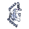
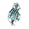
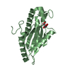
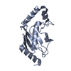
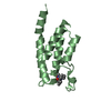
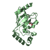
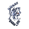
- PDB-6qh3: Catalytic domain of the human ubiquitin-conjugating enzyme UBE2S C118M -

+
Open data


ID or keywords:

Loading...

-
Basic information

Entry
Database: PDB / ID: 6qh3
TitleCatalytic domain of the human ubiquitin-conjugating enzyme UBE2S C118M
ComponentsUbiquitin-conjugating enzyme E2 S
KeywordsTRANSFERASE / human E2 / catalytic domain / C118M
Function / homology
Function and homology information


protein K29-linked ubiquitination / protein K27-linked ubiquitination / free ubiquitin chain polymerization / Conversion from APC/C:Cdc20 to APC/C:Cdh1 in late anaphase / Inactivation of APC/C via direct inhibition of the APC/C complex / APC/C:Cdc20 mediated degradation of mitotic proteins / anaphase-promoting complex / Aberrant regulation of mitotic exit in cancer due to RB1 defects / anaphase-promoting complex-dependent catabolic process / Phosphorylation of the APC/C ...protein K29-linked ubiquitination / protein K27-linked ubiquitination / free ubiquitin chain polymerization / Conversion from APC/C:Cdc20 to APC/C:Cdh1 in late anaphase / Inactivation of APC/C via direct inhibition of the APC/C complex / APC/C:Cdc20 mediated degradation of mitotic proteins / anaphase-promoting complex / Aberrant regulation of mitotic exit in cancer due to RB1 defects / anaphase-promoting complex-dependent catabolic process / Phosphorylation of the APC/C / anaphase-promoting complex binding / protein K6-linked ubiquitination / positive regulation of ubiquitin protein ligase activity / exit from mitosis / protein K11-linked ubiquitination / E2 ubiquitin-conjugating enzyme / ubiquitin conjugating enzyme activity / protein K63-linked ubiquitination / Regulation of APC/C activators between G1/S and early anaphase / Transcriptional Regulation by VENTX / APC/C:Cdc20 mediated degradation of Cyclin B / APC-Cdc20 mediated degradation of Nek2A / Synthesis of active ubiquitin: roles of E1 and E2 enzymes / Autodegradation of Cdh1 by Cdh1:APC/C / APC/C:Cdc20 mediated degradation of Securin / Assembly of the pre-replicative complex / Cdc20:Phospho-APC/C mediated degradation of Cyclin A / protein modification process / APC/C:Cdh1 mediated degradation of Cdc20 and other APC/C:Cdh1 targeted proteins in late mitosis/early G1 / CDK-mediated phosphorylation and removal of Cdc6 / protein polyubiquitination / ubiquitin-protein transferase activity / Separation of Sister Chromatids / Antigen processing: Ubiquitination & Proteasome degradation / ubiquitin-dependent protein catabolic process / Senescence-Associated Secretory Phenotype (SASP) / cell division / nucleoplasm / ATP binding / nucleus / cytosol
Similarity search - Function
Ubiquitin-conjugating enzyme, active site / Ubiquitin-conjugating (UBC) active site signature. / Ubiquitin-conjugating enzyme E2, catalytic domain homologues / Ubiquitin-conjugating enzyme E2 / Ubiquitin-conjugating enzyme / Ubiquitin-conjugating (UBC) core domain profile. / Ubiquitin-conjugating enzyme/RWD-like
Similarity search - Domain/homology
Ubiquitin-conjugating enzyme E2 S
Similarity search - Component
Biological speciesHomo sapiens (human)
MethodX-RAY DIFFRACTION / SYNCHROTRON / MOLECULAR REPLACEMENT / Resolution: 2.9 Å
AuthorsLiess, A.K.L. / Lorenz, S.
Funding support Germany, 2items
OrganizationGrant numberCountry
German Research FoundationEmmy Noether LO2003/1-1 Germany
German Research FoundationGRK 2243
CitationJournal: Structure / Year: 2019
Title: Autoinhibition Mechanism of the Ubiquitin-Conjugating Enzyme UBE2S by Autoubiquitination.
Authors: Liess, A.K.L. / Kucerova, A. / Schweimer, K. / Yu, L. / Roumeliotis, T.I. / Diebold, M. / Dybkov, O. / Sotriffer, C. / Urlaub, H. / Choudhary, J.S. / Mansfeld, J. / Lorenz, S.
History
DepositionJan 15, 2019Deposition site: PDBE / Processing site: PDBE
Revision 1.0Jul 17, 2019Provider: repository / Type: Initial release
Revision 1.1Aug 14, 2019Group: Data collection / Database references / Category: citation
Item: _citation.journal_volume / _citation.page_first / _citation.page_last
Revision 1.2Jan 24, 2024Group: Data collection / Database references / Refinement description
Category: chem_comp_atom / chem_comp_bond ...chem_comp_atom / chem_comp_bond / database_2 / pdbx_initial_refinement_model
Item: _database_2.pdbx_DOI / _database_2.pdbx_database_accession

-
Structure visualization

Structure viewerMolecule:
MolmilJmol/JSmol

Downloads & links

-
Assembly

Deposited unit
B: Ubiquitin-conjugating enzyme E2 S
A: Ubiquitin-conjugating enzyme E2 S
hetero molecules


Theoretical massNumber of molelcules
Total (without water)35,0826
Polymers34,8342
Non-polymers2484
Water0
1
B: Ubiquitin-conjugating enzyme E2 S
hetero molecules


Theoretical massNumber of molelcules
Total (without water)17,6034
Polymers17,4171
Non-polymers1863
Water0
TypeNameSymmetry operationNumber
identity operation1_555x,y,z1
2
A: Ubiquitin-conjugating enzyme E2 S
hetero molecules


Theoretical massNumber of molelcules
Total (without water)17,4792
Polymers17,4171
Non-polymers621
Water0
TypeNameSymmetry operationNumber
identity operation1_555x,y,z1
Unit cell
Length a, b, c (Å)84.619, 84.619, 87.832
Angle α, β, γ (deg.)90.00, 90.00, 120.00
Int Tables number170
Space group name H-MP65

-
Components

#1: Protein Ubiquitin-conjugating enzyme E2 S / E2-EPF / Ubiquitin carrier protein S / Ubiquitin-conjugating enzyme E2-24 kDa / Ubiquitin- ...E2-EPF / Ubiquitin carrier protein S / Ubiquitin-conjugating enzyme E2-24 kDa / Ubiquitin-conjugating enzyme E2-EPF5 / Ubiquitin-protein ligase S


Mass: 17417.012 Da / Num. of mol.: 2 / Mutation: C118M
Source method: isolated from a genetically manipulated source
Source: (gene. exp.) Homo sapiens (human) / Gene: UBE2S, E2EPF, OK/SW-cl.73 / Production host: Escherichia coli BL21(DE3) (bacteria)
References: UniProt: Q16763, E2 ubiquitin-conjugating enzyme
#2: Chemical
ChemComp-EDO / 1,2-ETHANEDIOL / ETHYLENE GLYCOL / Ethylene glycol


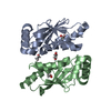
Mass: 62.068 Da / Num. of mol.: 4 / Source method: obtained synthetically / Formula: C2H6O2

-
Experimental details

-
Experiment

ExperimentMethod: X-RAY DIFFRACTION / Number of used crystals: 1

-
Sample preparation

CrystalDensity Matthews: 2.61 Å3/Da / Density % sol: 52.8 %
Crystal growTemperature: 293 K / Method: vapor diffusion, sitting drop / pH: 7.5
Details: 0.2 M magnesium chloride, 0.1 M hepes pH 7.5, 25% PEG 3350

-
Data collection

DiffractionMean temperature: 100 K / Serial crystal experiment: N
Diffraction sourceSource: SYNCHROTRON / Site: PETRA III, EMBL c/o DESY / Beamline: P14 (MX2) / Wavelength: 0.9762 Å
DetectorType: DECTRIS PILATUS 6M-F / Detector: PIXEL / Date: Dec 1, 2016
RadiationProtocol: SINGLE WAVELENGTH / Monochromatic (M) / Laue (L): M / Scattering type: x-ray
Radiation wavelengthWavelength: 0.9762 Å / Relative weight: 1
ReflectionResolution: 2.9→42.309 Å / Num. obs: 7984 / % possible obs: 99.94 % / Redundancy: 2 % / Biso Wilson estimate: 63.58 Å2 / CC1/2: 0.999 / Rmerge(I) obs: 0.02634 / Rpim(I) all: 0.02634 / Rrim(I) all: 0.03725 / Net I/σ(I): 21.4
Reflection shellResolution: 2.9→3.004 Å / Redundancy: 2 % / Rmerge(I) obs: 0.1729 / Mean I/σ(I) obs: 4.19 / Num. unique obs: 795 / CC1/2: 0.896 / Rpim(I) all: 0.1729 / Rrim(I) all: 0.2445 / % possible all: 100

-
Processing

Software
NameVersionClassification
PHENIX(1.11_2567: ???)refinement
XDSdata reduction
Aimlessdata scaling
PHASERphasing
RefinementMethod to determine structure: MOLECULAR REPLACEMENT
Starting model: 1ZDN
Resolution: 2.9→42.309 Å / SU ML: 0.35 / Cross valid method: FREE R-VALUE / σ(F): 1.38 / Phase error: 26.93
RfactorNum. reflection% reflection
Rfree0.23 439 5.5 %
Rwork0.1843 --
obs0.187 7984 99.97 %
Solvent computationShrinkage radii: 0.9 Å / VDW probe radii: 1.11 Å
Refinement stepCycle: LAST / Resolution: 2.9→42.309 Å
ProteinNucleic acidLigandSolventTotal
Num. atoms2254 0 16 0 2270
Refine LS restraints
Refine-IDTypeDev idealNumber
X-RAY DIFFRACTIONf_bond_d0.0032323
X-RAY DIFFRACTIONf_angle_d0.7043163
X-RAY DIFFRACTIONf_dihedral_angle_d15.8941390
X-RAY DIFFRACTIONf_chiral_restr0.044365
X-RAY DIFFRACTIONf_plane_restr0.005408
LS refinement shell
Resolution (Å)Rfactor RfreeNum. reflection RfreeRfactor RworkNum. reflection RworkRefine-ID% reflection obs (%)
2.9001-3.31960.29221130.21742518X-RAY DIFFRACTION100
3.3196-4.18180.22621770.19192495X-RAY DIFFRACTION100
4.1818-42.3090.21851490.16962532X-RAY DIFFRACTION100
Refinement TLS params.

Method: refined / Refine-ID: X-RAY DIFFRACTION

IDL112)L122)L132)L222)L232)L332)S11 (Å °)S12 (Å °)S13 (Å °)S21 (Å °)S22 (Å °)S23 (Å °)S31 (Å °)S32 (Å °)S33 (Å °)T112)T122)T132)T222)T232)T332)Origin x (Å)Origin y (Å)Origin z (Å)
15.91650.9954-1.13517.98040.04445.4084-0.37470.3813-0.3574-0.48790.2177-0.32260.2547-0.00120.11350.3076-0.14320.04640.3955-0.00580.4029120.3938-37.8424-4.8241
24.7323-0.93380.94617.6026-2.84015.9694-0.1009-0.01690.10520.1458-0.05210.18630.0395-0.43280.16180.2267-0.10310.06590.4997-0.02780.331299.2848-43.61811.8454
Refinement TLS group
IDRefine-IDRefine TLS-IDSelection details
1X-RAY DIFFRACTION1(chain 'B' and resid 10 through 156)
2X-RAY DIFFRACTION2(chain 'A' and resid 9 through 156)

+
About Yorodumi

-
News

-
Feb 9, 2022. New format data for meta-information of EMDB entries

New format data for meta-information of EMDB entries

  • Version 3 of the EMDB header file is now the official format.
  • The previous official version 1.9 will be removed from the archive.

Related info.:EMDB header

External links:wwPDB to switch to version 3 of the EMDB data model

-
Aug 12, 2020. Covid-19 info

Covid-19 info

URL: https://pdbj.org/emnavi/covid19.php

New page: Covid-19 featured information page in EM Navigator.

Related info.:Covid-19 info / Mar 5, 2020. Novel coronavirus structure data

+
Mar 5, 2020. Novel coronavirus structure data

Novel coronavirus structure data

Related info.:Yorodumi Speices / Aug 12, 2020. Covid-19 info

External links:COVID-19 featured content - PDBj / Molecule of the Month (242):Coronavirus Proteases

+
Jan 31, 2019. EMDB accession codes are about to change! (news from PDBe EMDB page)

EMDB accession codes are about to change! (news from PDBe EMDB page)

  • The allocation of 4 digits for EMDB accession codes will soon come to an end. Whilst these codes will remain in use, new EMDB accession codes will include an additional digit and will expand incrementally as the available range of codes is exhausted. The current 4-digit format prefixed with “EMD-” (i.e. EMD-XXXX) will advance to a 5-digit format (i.e. EMD-XXXXX), and so on. It is currently estimated that the 4-digit codes will be depleted around Spring 2019, at which point the 5-digit format will come into force.
  • The EM Navigator/Yorodumi systems omit the EMD- prefix.

Related info.:Q: What is EMD? / ID/Accession-code notation in Yorodumi/EM Navigator

External links:EMDB Accession Codes are Changing Soon! / Contact to PDBj

+
Jul 12, 2017. Major update of PDB

Major update of PDB

  • wwPDB released updated PDB data conforming to the new PDBx/mmCIF dictionary.
  • This is a major update changing the version number from 4 to 5, and with Remediation, in which all the entries are updated.
  • In this update, many items about electron microscopy experimental information are reorganized (e.g. em_software).
  • Now, EM Navigator and Yorodumi are based on the updated data.

External links:wwPDB Remediation / Enriched Model Files Conforming to OneDep Data Standards Now Available in the PDB FTP Archive

-
Yorodumi

Thousand views of thousand structures

  • Yorodumi is a browser for structure data from EMDB, PDB, SASBDB, etc.
  • This page is also the successor to EM Navigator detail page, and also detail information page/front-end page for Omokage search.
  • The word "yorodu" (or yorozu) is an old Japanese word meaning "ten thousand". "mi" (miru) is to see.

Related info.:EMDB / PDB / SASBDB / Comparison of 3 databanks / Yorodumi Search / Aug 31, 2016. New EM Navigator & Yorodumi / Yorodumi Papers / Jmol/JSmol / Function and homology information / Changes in new EM Navigator and Yorodumi

Read more