 Movie
Movie Controller

[English] 日本語
 Yorodumi
- PDB-6q57: X-ray crystal structure of the tetrahydrofolate riboswitch aptame... -
+ Open data

- Basic information
| Entry | Database: PDB / ID: 6q57 | ||||||
|---|---|---|---|---|---|---|---|
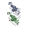
| Title | X-ray crystal structure of the tetrahydrofolate riboswitch aptamer bound to 5-deazatetrahydropterin | ||||||
 Components Components | tetrahydrofolate riboswitch aptamer | ||||||
 Keywords Keywords | RNA / Aptamer / riboswitch | ||||||
| Function / homology | 5-deazatetrahydropterin / RNA / RNA (> 10) Function and homology information Function and homology information | ||||||
| Biological species |  Streptococcus mutans UA159 (bacteria) Streptococcus mutans UA159 (bacteria) | ||||||
| Method |  X-RAY DIFFRACTION / SYNCHROTRON / Resolution: 2.72 Å X-RAY DIFFRACTION / SYNCHROTRON / Resolution: 2.72 Å | ||||||
 Authors Authors | Dunstan, M.S. | ||||||
| Funding support |  United Kingdom, 1items United Kingdom, 1items
| ||||||
 Citation Citation | Journal: To Be Published Title: Tetrahydrofolate Riboswitches Provide Distinct Genetic Outputs to Synthetic and Natural Signals. Authors: Vincent, H.A. / Leigh, J. / Robinson, C.J. / Dunstan, M.S. / Ferrer Rios, M.G. / Micklefield, J. | ||||||
| History |
|
- Structure visualization
| Structure viewer | Molecule: MolmilJmol/JSmol |
|---|
- Downloads & links
-Download
| PDBx/mmCIF format | 6q57.cif.gz | 60.2 KB | Display | PDBx/mmCIF format |
|---|---|---|---|---|
| PDB format | pdb6q57.ent.gz | 42.7 KB | Display | PDB format |
| PDBx/mmJSON format | 6q57.json.gz | Tree view | PDBx/mmJSON format | |
| Others |  Other downloads Other downloads |
-Validation report
| Arichive directory | https://data.pdbj.org/pub/pdb/validation_reports/q5/6q57ftp://data.pdbj.org/pub/pdb/validation_reports/q5/6q57 | HTTPS FTP |
|---|
-Related structure data
| Similar structure data |
|---|


-Links
 PDBj
PDBj


- Assembly
| Deposited unit | 
| ||||||||
|---|---|---|---|---|---|---|---|---|---|
| 1 |
| ||||||||
| Unit cell |
|
-Components
| #1: RNA chain | Mass: 28849.145 Da / Num. of mol.: 1 Source method: isolated from a genetically manipulated source Source: (gene. exp.) Streptococcus mutans UA159 (bacteria) / Production host: | ||||
|---|---|---|---|---|---|
| #2: Chemical |   Mass: 166.180 Da / Num. of mol.: 2 / Source method: obtained synthetically / Formula: C7H10N4O Mass: 166.180 Da / Num. of mol.: 2 / Source method: obtained synthetically / Formula: C7H10N4O#3: Chemical | ChemComp-MG / |   Mass: 24.305 Da / Num. of mol.: 1 / Source method: obtained synthetically / Formula: Mg Mass: 24.305 Da / Num. of mol.: 1 / Source method: obtained synthetically / Formula: Mg#4: Water | ChemComp-HOH / |  Mass: 18.015 Da / Num. of mol.: 2 / Source method: isolated from a natural source / Formula: H2O Mass: 18.015 Da / Num. of mol.: 2 / Source method: isolated from a natural source / Formula: H2O |
-Experimental details
-Experiment
| Experiment | Method: X-RAY DIFFRACTION / Number of used crystals: 1 |
|---|
- Sample preparation
| Crystal | Density Matthews: 2.52 Å3/Da / Density % sol: 51.11 % |
|---|---|
| Crystal grow | Temperature: 303 K / Method: vapor diffusion, sitting drop Details: 6% 2,4-methyl-pentanediol, 10 mM spermine, 40 mM Na-cacodylate, pH 7.0, 80 mM NaCl and 1 mM DTT |
-Data collection
| Diffraction | Mean temperature: 173 K / Serial crystal experiment: N |
|---|---|
| Diffraction source | Source: SYNCHROTRON / Site: Diamond / Beamline: I23 / Wavelength: 1 Å |
| Detector | Type: DECTRIS PILATUS3 S 6M / Detector: PIXEL / Date: Dec 18, 2015 |
| Radiation | Protocol: SINGLE WAVELENGTH / Monochromatic (M) / Laue (L): M / Scattering type: x-ray |
| Radiation wavelength | Wavelength: 1 Å / Relative weight: 1 |
| Reflection | Resolution: 2.72→51.741 Å / Num. obs: 8282 / % possible obs: 98 % / Redundancy: 6.1 % / Net I/σ(I): 6.7 |
| Reflection shell | Resolution: 2.72→2.82 Å |
- Processing
| Software |
| ||||||||||||||||||||||||||||
|---|---|---|---|---|---|---|---|---|---|---|---|---|---|---|---|---|---|---|---|---|---|---|---|---|---|---|---|---|---|
| Refinement | Resolution: 2.72→51.741 Å / SU ML: 0.5 / Cross valid method: THROUGHOUT / σ(F): 1.35 / Phase error: 36.24
| ||||||||||||||||||||||||||||
| Solvent computation | Shrinkage radii: 0.9 Å / VDW probe radii: 1.11 Å | ||||||||||||||||||||||||||||
| Displacement parameters | Biso max: 120.84 Å2 / Biso mean: 60.1665 Å2 / Biso min: 25.48 Å2 | ||||||||||||||||||||||||||||
| Refinement step | Cycle: final / Resolution: 2.72→51.741 Å
| ||||||||||||||||||||||||||||
| Refine LS restraints |
| ||||||||||||||||||||||||||||
| LS refinement shell | Refine-ID: X-RAY DIFFRACTION / Rfactor Rfree error: 0 / Total num. of bins used: 3
|
