 Movie
Movie Controller

[English] 日本語
 Yorodumi
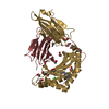
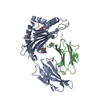
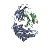
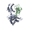
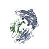
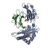
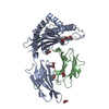
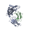
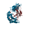
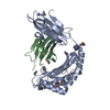
- PDB-6pyl: Crystal Structure of HLA-B*2703 in complex with KK10, an HIV peptide -
+ Open data

- Basic information
| Entry | Database: PDB / ID: 6pyl | ||||||
|---|---|---|---|---|---|---|---|
| Title | Crystal Structure of HLA-B*2703 in complex with KK10, an HIV peptide | ||||||
 Components Components |
| ||||||
 Keywords Keywords | IMMUNE SYSTEM / Ankylosing spondylitis / HLA-B27 / HLA-B*27:03 / HLA-B*27:05 / HLA | ||||||
| Function / homology |  Function and homology information Function and homology informationregulation of interleukin-12 production / regulation of dendritic cell differentiation / integrase activity / regulation of T cell anergy / regulation of interleukin-6 production / Integration of viral DNA into host genomic DNA / Autointegration results in viral DNA circles / Minus-strand DNA synthesis / Plus-strand DNA synthesis / Uncoating of the HIV Virion ...regulation of interleukin-12 production / regulation of dendritic cell differentiation / integrase activity / regulation of T cell anergy / regulation of interleukin-6 production / Integration of viral DNA into host genomic DNA / Autointegration results in viral DNA circles / Minus-strand DNA synthesis / Plus-strand DNA synthesis / Uncoating of the HIV Virion / 2-LTR circle formation / Vpr-mediated nuclear import of PICs / Early Phase of HIV Life Cycle / Integration of provirus / APOBEC3G mediated resistance to HIV-1 infection / protection from natural killer cell mediated cytotoxicity / TAP binding / Binding and entry of HIV virion / detection of bacterium / antigen processing and presentation of endogenous peptide antigen via MHC class Ib / antigen processing and presentation of endogenous peptide antigen via MHC class I via ER pathway, TAP-independent / viral life cycle / secretory granule membrane / early endosome lumen / Nef mediated downregulation of MHC class I complex cell surface expression / DAP12 interactions / Endosomal/Vacuolar pathway / T cell mediated cytotoxicity / HIV-1 retropepsin / symbiont-mediated activation of host apoptosis / retroviral ribonuclease H / exoribonuclease H / Antigen Presentation: Folding, assembly and peptide loading of class I MHC / lumenal side of endoplasmic reticulum membrane / regulation of iron ion transport / cellular response to iron(III) ion / negative regulation of iron ion transport / negative regulation of forebrain neuron differentiation / antigen processing and presentation of exogenous protein antigen via MHC class Ib, TAP-dependent / exoribonuclease H activity / peptide antigen assembly with MHC class I protein complex / ER to Golgi transport vesicle membrane / regulation of erythrocyte differentiation / response to molecule of bacterial origin / HFE-transferrin receptor complex / defense response / MHC class I peptide loading complex / transferrin transport / cellular response to iron ion / negative regulation of receptor-mediated endocytosis / Assembly Of The HIV Virion / positive regulation of T cell cytokine production / antigen processing and presentation of endogenous peptide antigen via MHC class I / MHC class I protein complex / protein processing / peptide antigen assembly with MHC class II protein complex / Budding and maturation of HIV virion / viral genome integration into host DNA / negative regulation of neurogenesis / cellular response to nicotine / MHC class II protein complex / positive regulation of receptor-mediated endocytosis / establishment of integrated proviral latency / RNA-directed DNA polymerase / multicellular organismal-level iron ion homeostasis / positive regulation of T cell mediated cytotoxicity / RNA stem-loop binding / viral penetration into host nucleus / specific granule lumen / antigen processing and presentation of exogenous peptide antigen via MHC class II / positive regulation of immune response / host multivesicular body / peptide antigen binding / RNA-directed DNA polymerase activity / phagocytic vesicle membrane / RNA-DNA hybrid ribonuclease activity / recycling endosome membrane / positive regulation of T cell activation / Interferon gamma signaling / negative regulation of epithelial cell proliferation / Immunoregulatory interactions between a Lymphoid and a non-Lymphoid cell / Transferases; Transferring phosphorus-containing groups; Nucleotidyltransferases / Interferon alpha/beta signaling / Modulation by Mtb of host immune system / sensory perception of smell / positive regulation of cellular senescence / tertiary granule lumen / MHC class II protein complex binding / T cell differentiation in thymus / DAP12 signaling / peptidase activity / late endosome membrane / host cell / negative regulation of neuron projection development / protein refolding / protein-folding chaperone binding / viral nucleocapsid / ER-Phagosome pathway / DNA recombination / early endosome membrane Similarity search - Function | ||||||
| Biological species |  Homo sapiens (human) Homo sapiens (human)  Human immunodeficiency virus 1 Human immunodeficiency virus 1 | ||||||
| Method |  X-RAY DIFFRACTION / SYNCHROTRON / MOLECULAR REPLACEMENT / molecular replacement / Resolution: 1.52 Å X-RAY DIFFRACTION / SYNCHROTRON / MOLECULAR REPLACEMENT / molecular replacement / Resolution: 1.52 Å | ||||||
 Authors Authors | Gras, S. | ||||||
 Citation Citation | Journal: J.Biol.Chem. / Year: 2019 Title: Allelic association with ankylosing spondylitis fails to correlate with human leukocyte antigen B27 homodimer formation. Authors: Lim Kam Sian, T.C.C. / Indumathy, S. / Halim, H. / Greule, A. / Cryle, M.J. / Bowness, P. / Rossjohn, J. / Gras, S. / Purcell, A.W. / Schittenhelm, R.B. | ||||||
| History |
|
- Structure visualization
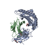
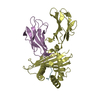
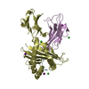
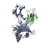
| Structure viewer | Molecule: MolmilJmol/JSmol |
|---|
- Downloads & links
-Download
| PDBx/mmCIF format | 6pyl.cif.gz | 105.8 KB | Display | PDBx/mmCIF format |
|---|---|---|---|---|
| PDB format | pdb6pyl.ent.gz | 77.5 KB | Display | PDB format |
| PDBx/mmJSON format | 6pyl.json.gz | Tree view | PDBx/mmJSON format | |
| Others |  Other downloads Other downloads |
-Validation report
| Arichive directory | https://data.pdbj.org/pub/pdb/validation_reports/py/6pylftp://data.pdbj.org/pub/pdb/validation_reports/py/6pyl | HTTPS FTP |
|---|
-Related structure data
| Related structure data |  6pyjC 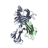 6pyvC  6pywC  6pz5C  4g9dS S: Starting model for refinement C: citing same article ( |
|---|---|
| Similar structure data |


-Links
 PDBj
PDBj


- Assembly
| Deposited unit | 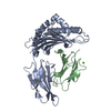
| ||||||||
|---|---|---|---|---|---|---|---|---|---|
| 1 |
| ||||||||
| Unit cell |
|
-Components
| #1: Protein | Mass: 31903.133 Da / Num. of mol.: 1 Source method: isolated from a genetically manipulated source Source: (gene. exp.) Homo sapiens (human) / Gene: HLA-B, HLAB / Production host:  |
|---|---|
| #2: Protein | Mass: 11879.356 Da / Num. of mol.: 1 Source method: isolated from a genetically manipulated source Source: (gene. exp.) Homo sapiens (human) / Gene: B2M, CDABP0092, HDCMA22P / Production host: |
| #3: Protein/peptide | Mass: 1243.564 Da / Num. of mol.: 1 / Source method: obtained synthetically / Source: (synth.) Human immunodeficiency virus 1 / References: UniProt: P04585*PLUS |
| #4: Chemical | ChemComp-MG /   Mass: 24.305 Da / Num. of mol.: 1 / Source method: obtained synthetically / Formula: Mg Mass: 24.305 Da / Num. of mol.: 1 / Source method: obtained synthetically / Formula: Mg |
| #5: Water | ChemComp-HOH /  Mass: 18.015 Da / Num. of mol.: 423 / Source method: isolated from a natural source / Formula: H2O Mass: 18.015 Da / Num. of mol.: 423 / Source method: isolated from a natural source / Formula: H2O |
| Has ligand of interest | N |
| Has protein modification | Y |
| Sequence details | Y83H in allele B*27:03 |
-Experimental details
-Experiment
| Experiment | Method: X-RAY DIFFRACTION / Number of used crystals: 1 |
|---|
- Sample preparation
| Crystal | Density Matthews: 2.44 Å3/Da / Density % sol: 49.68 % |
|---|---|
| Crystal grow | Temperature: 277 K / Method: evaporation Details: 20-30% PEG 4K, 0.2M Na Acetate and 0.1M Na Citrate pH 5.6 |
-Data collection
| Diffraction | Mean temperature: 100 K / Serial crystal experiment: N |
|---|---|
| Diffraction source | Source: SYNCHROTRON / Site: Australian Synchrotron  / Beamline: MX2 / Wavelength: 0.9537 Å / Beamline: MX2 / Wavelength: 0.9537 Å |
| Detector | Type: ADSC QUANTUM 315r / Detector: CCD / Date: Aug 14, 2015 |
| Radiation | Protocol: SINGLE WAVELENGTH / Monochromatic (M) / Laue (L): M / Scattering type: x-ray |
| Radiation wavelength | Wavelength: 0.9537 Å / Relative weight: 1 |
| Reflection | Resolution: 1.52→44.24 Å / Num. obs: 68648 / % possible obs: 100 % / Redundancy: 7.3 % / Biso Wilson estimate: 16.34 Å2 / Rpim(I) all: 0.04 / Net I/σ(I): 14.5 |
| Reflection shell | Resolution: 1.52→1.55 Å / Mean I/σ(I) obs: 2.1 / Num. unique obs: 24151 / Rpim(I) all: 0.423 |
-Phasing
| Phasing | Method: molecular replacement |
|---|
- Processing
| Software |
| ||||||||||||||||||||||||||||||||||||||||||||||||||||||||||||||||||||||||||||||||||||||||||||||||||||||||||||
|---|---|---|---|---|---|---|---|---|---|---|---|---|---|---|---|---|---|---|---|---|---|---|---|---|---|---|---|---|---|---|---|---|---|---|---|---|---|---|---|---|---|---|---|---|---|---|---|---|---|---|---|---|---|---|---|---|---|---|---|---|---|---|---|---|---|---|---|---|---|---|---|---|---|---|---|---|---|---|---|---|---|---|---|---|---|---|---|---|---|---|---|---|---|---|---|---|---|---|---|---|---|---|---|---|---|---|---|---|---|
| Refinement | Method to determine structure: MOLECULAR REPLACEMENT Starting model: 4g9d Resolution: 1.52→30.4 Å / Cor.coef. Fo:Fc: 0.923 / Cor.coef. Fo:Fc free: 0.907 / SU R Cruickshank DPI: 0.079 / Cross valid method: THROUGHOUT / σ(F): 0 / SU R Blow DPI: 0.084 / SU Rfree Blow DPI: 0.084 / SU Rfree Cruickshank DPI: 0.08
| ||||||||||||||||||||||||||||||||||||||||||||||||||||||||||||||||||||||||||||||||||||||||||||||||||||||||||||
| Displacement parameters | Biso max: 134.66 Å2 / Biso mean: 19.45 Å2 / Biso min: 5.79 Å2
| ||||||||||||||||||||||||||||||||||||||||||||||||||||||||||||||||||||||||||||||||||||||||||||||||||||||||||||
| Refine analyze | Luzzati coordinate error obs: 0.23 Å | ||||||||||||||||||||||||||||||||||||||||||||||||||||||||||||||||||||||||||||||||||||||||||||||||||||||||||||
| Refinement step | Cycle: final / Resolution: 1.52→30.4 Å
| ||||||||||||||||||||||||||||||||||||||||||||||||||||||||||||||||||||||||||||||||||||||||||||||||||||||||||||
| Refine LS restraints |
| ||||||||||||||||||||||||||||||||||||||||||||||||||||||||||||||||||||||||||||||||||||||||||||||||||||||||||||
| LS refinement shell | Resolution: 1.52→1.56 Å / Rfactor Rfree error: 0 / Total num. of bins used: 20
|
