[English] 日本語
Yorodumi
- PDB-6pqa: GAVVGG segment 119-124 from human prion -

+
Open data


ID or keywords:

Loading...

-
Basic information

Entry
Database: PDB / ID: 6pqa
TitleGAVVGG segment 119-124 from human prion
ComponentsMajor prion protein
KeywordsPROTEIN FIBRIL / amyloid-like protofibril
Function / homology
Function and homology information


positive regulation of glutamate receptor signaling pathway / negative regulation of amyloid precursor protein catabolic process / lamin binding / regulation of glutamate receptor signaling pathway / regulation of calcium ion import across plasma membrane / aspartic-type endopeptidase inhibitor activity / glycosaminoglycan binding / ATP-dependent protein binding / regulation of potassium ion transmembrane transport / negative regulation of interleukin-17 production ...positive regulation of glutamate receptor signaling pathway / negative regulation of amyloid precursor protein catabolic process / lamin binding / regulation of glutamate receptor signaling pathway / regulation of calcium ion import across plasma membrane / aspartic-type endopeptidase inhibitor activity / glycosaminoglycan binding / ATP-dependent protein binding / regulation of potassium ion transmembrane transport / negative regulation of interleukin-17 production / NCAM1 interactions / negative regulation of dendritic spine maintenance / type 5 metabotropic glutamate receptor binding / cupric ion binding / negative regulation of protein processing / negative regulation of calcineurin-NFAT signaling cascade / dendritic spine maintenance / negative regulation of interleukin-2 production / negative regulation of T cell receptor signaling pathway / Insertion of tail-anchored proteins into the endoplasmic reticulum membrane / extrinsic component of membrane / cuprous ion binding / negative regulation of amyloid-beta formation / negative regulation of activated T cell proliferation / response to amyloid-beta / : / negative regulation of type II interferon production / negative regulation of long-term synaptic potentiation / intracellular copper ion homeostasis / positive regulation of protein targeting to membrane / long-term memory / response to cadmium ion / regulation of peptidyl-tyrosine phosphorylation / inclusion body / cellular response to copper ion / neuron projection maintenance / tubulin binding / negative regulation of protein phosphorylation / molecular condensate scaffold activity / molecular function activator activity / positive regulation of protein localization to plasma membrane / protein destabilization / protein homooligomerization / negative regulation of DNA-binding transcription factor activity / terminal bouton / cellular response to amyloid-beta / positive regulation of peptidyl-tyrosine phosphorylation / positive regulation of neuron apoptotic process / cellular response to xenobiotic stimulus / signaling receptor activity / amyloid-beta binding / protein-folding chaperone binding / postsynapse / microtubule binding / nuclear membrane / protease binding / response to oxidative stress / transmembrane transporter binding / molecular adaptor activity / postsynaptic density / learning or memory / regulation of cell cycle / membrane raft / cell cycle / copper ion binding / external side of plasma membrane / intracellular membrane-bounded organelle / dendrite / protein-containing complex binding / negative regulation of apoptotic process / Golgi apparatus / cell surface / endoplasmic reticulum / extracellular exosome / identical protein binding / plasma membrane / cytoplasm / cytosol
Similarity search - Function
Prion protein signature 1. / Prion protein signature 2. / Major prion protein N-terminal domain / Major prion protein bPrPp - N terminal / Prion protein / Major prion protein / Prion/Doppel protein, beta-ribbon domain / Prion/Doppel beta-ribbon domain superfamily / Prion/Doppel alpha-helical domain
Similarity search - Domain/homology
Biological speciesHomo sapiens (human)
MethodX-RAY DIFFRACTION / SYNCHROTRON / MOLECULAR REPLACEMENT / molecular replacement / Resolution: 1.46 Å
AuthorsApostol, M.I. / Sawaya, M.R. / Eisenberg, D.
Funding support United States, 4items
OrganizationGrant numberCountry
National Institutes of Health/National Institute on Aging (NIH/NIA)R01 AG029430 United States
National Institutes of Health/National Institute on Aging (NIH/NIA)R01 AG029430-05 United States
National Institutes of Health/National Institute of General Medical Sciences (NIH/NIGMS)T32 GM007185 United States
National Institutes of Health/National Institute of General Medical Sciences (NIH/NIGMS)GM007185 United States
Citation
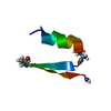
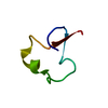
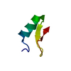
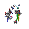
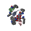
Journal: Nat Struct Mol Biol / Year: 2020
Title: Cryo-EM structure of a human prion fibril with a hydrophobic, protease-resistant core.
Authors: Calina Glynn / Michael R Sawaya / Peng Ge / Marcus Gallagher-Jones / Connor W Short / Ronquiajah Bowman / Marcin Apostol / Z Hong Zhou / David S Eisenberg / Jose A Rodriguez /
Abstract: Self-templating assemblies of the human prion protein are clinically associated with transmissible spongiform encephalopathies. Here we present the cryo-EM structure of a denaturant- and protease- ...Self-templating assemblies of the human prion protein are clinically associated with transmissible spongiform encephalopathies. Here we present the cryo-EM structure of a denaturant- and protease-resistant fibril formed in vitro spontaneously by a 9.7-kDa unglycosylated fragment of the human prion protein. This human prion fibril contains two protofilaments intertwined with screw symmetry and linked by a tightly packed hydrophobic interface. Each protofilament consists of an extended beta arch formed by residues 106 to 145 of the prion protein, a hydrophobic and highly fibrillogenic disease-associated segment. Such structures of prion polymorphs serve as blueprints on which to evaluate the potential impact of sequence variants on prion disease.
History
DepositionJul 8, 2019Deposition site: RCSB / Processing site: RCSB
Revision 1.0Apr 15, 2020Provider: repository / Type: Initial release
Revision 1.1Apr 29, 2020Group: Database references / Category: citation / citation_author
Item: _citation.pdbx_database_id_PubMed / _citation.title / _citation_author.name
Revision 1.2May 27, 2020Group: Database references / Category: citation
Item: _citation.journal_volume / _citation.page_first / _citation.page_last
Revision 1.3Mar 13, 2024Group: Data collection / Database references / Category: chem_comp_atom / chem_comp_bond / database_2
Item: _database_2.pdbx_DOI / _database_2.pdbx_database_accession

-
Structure visualization

Structure viewerMolecule:
MolmilJmol/JSmol

Downloads & links

-
Assembly

Deposited unit
A: Major prion protein


Theoretical massNumber of molelcules
Total (without water)4591
Polymers4591
Non-polymers00
Water543
1
A: Major prion protein

A: Major prion protein

A: Major prion protein

A: Major prion protein

A: Major prion protein

A: Major prion protein

A: Major prion protein

A: Major prion protein


Theoretical massNumber of molelcules
Total (without water)3,6688
Polymers3,6688
Non-polymers00
Water1448
TypeNameSymmetry operationNumber
identity operation1_555x,y,z1
crystal symmetry operation1_655x+1,y,z1
crystal symmetry operation1_755x+2,y,z1
crystal symmetry operation1_855x+3,y,z1
crystal symmetry operation2_554-x,y+1/2,-z-11
crystal symmetry operation2_654-x+1,y+1/2,-z-11
crystal symmetry operation2_754-x+2,y+1/2,-z-11
crystal symmetry operation2_854-x+3,y+1/2,-z-11
Unit cell
Length a, b, c (Å)4.786, 12.810, 20.771
Angle α, β, γ (deg.)90.000, 92.380, 90.000
Int Tables number4
Space group name H-MP1211

-
Components

#1: Protein/peptide Major prion protein / PrP / ASCR / PrP27-30 / PrP33-35C


Mass: 458.510 Da / Num. of mol.: 1 / Fragment: UNP residues 119-124 / Source method: obtained synthetically / Source: (synth.) Homo sapiens (human) / References: UniProt: P04156
#2: Water ChemComp-HOH / water


Mass: 18.015 Da / Num. of mol.: 3 / Source method: isolated from a natural source / Formula: H2O

-
Experimental details

-
Experiment

ExperimentMethod: X-RAY DIFFRACTION / Number of used crystals: 1

-
Sample preparation

CrystalDensity Matthews: 1.39 Å3/Da / Density % sol: 11.35 %
Crystal growTemperature: 298 K / Method: vapor diffusion, hanging drop / pH: 7 / Details: 10 mg/mL peptide in 2.4 M sodium malonate, pH 7.0 / PH range: 6.8-7.2

-
Data collection

DiffractionMean temperature: 100 K / Serial crystal experiment: N
Diffraction sourceSource: SYNCHROTRON / Site: ESRF / Beamline: ID13 / Wavelength: 0.9465 Å
DetectorType: MAR CCD 165 mm / Detector: CCD / Date: May 13, 2006
RadiationMonochromator: Si(111) / Protocol: SINGLE WAVELENGTH / Monochromatic (M) / Laue (L): M / Scattering type: x-ray
Radiation wavelengthWavelength: 0.9465 Å / Relative weight: 1
ReflectionResolution: 1.45→30 Å / Num. obs: 455 / % possible obs: 93.6 % / Redundancy: 5.4 % / Rmerge(I) obs: 0.145 / Rsym value: 0.15 / Χ2: 1.052 / Net I/σ(I): 31.4
Reflection shell

Diffraction-ID: 1

Resolution (Å)Redundancy (%)Rmerge(I) obsNum. unique obsRsym valueΧ2% possible all
1.45-1.52.80.311410.311.00693.2
1.5-1.563.70.274481.09984.2
1.56-1.634.50.187410.96280.4
1.63-1.726.10.218361.145100
1.72-1.836.50.242461.124100
1.83-1.976.10.137481.291100
1.97-2.176.20.143510.888100
2.17-2.485.90.152501.00394.3
2.48-3.126.40.132440.984100
3.12-305.40.095500.99889.3

-
Phasing

PhasingMethod: molecular replacement

-
Processing

Software
NameVersionClassification
DENZOdata reduction
SCALEPACK1.97.8data scaling
PHASER1.3.2phasing
REFMAC5.4.0061refinement
PDB_EXTRACT3.25data extraction
RefinementMethod to determine structure: MOLECULAR REPLACEMENT / Resolution: 1.46→20.75 Å / Cor.coef. Fo:Fc: 0.948 / Cor.coef. Fo:Fc free: 0.878 / SU B: 1.099 / SU ML: 0.044 / SU R Cruickshank DPI: 0.1047 / Cross valid method: THROUGHOUT / σ(F): 0 / ESU R: 0.105 / ESU R Free: 0.111 / Details: HYDROGENS HAVE BEEN ADDED IN THE RIDING POSITIONS
RfactorNum. reflection% reflectionSelection details
Rfree0.2379 46 10.6 %RANDOM
Rwork0.1802 ---
obs0.1867 389 92.75 %-
Solvent computationIon probe radii: 0.8 Å / Shrinkage radii: 0.8 Å / VDW probe radii: 1.4 Å
Displacement parametersBiso max: 33.98 Å2 / Biso mean: 4.425 Å2 / Biso min: 0.88 Å2
Baniso -1Baniso -2Baniso -3
1--0.3 Å20 Å2-0.28 Å2
2--0.88 Å20 Å2
3----0.6 Å2
Refinement stepCycle: final / Resolution: 1.46→20.75 Å
ProteinNucleic acidLigandSolventTotal
Num. atoms32 0 0 3 35
Biso mean---31.46 -
Num. residues----6
Refine LS restraints
Refine-IDTypeDev idealDev ideal targetNumber
X-RAY DIFFRACTIONr_bond_refined_d0.0070.02231
X-RAY DIFFRACTIONr_bond_other_d0.0010.0216
X-RAY DIFFRACTIONr_angle_refined_deg1.0812.03941
X-RAY DIFFRACTIONr_angle_other_deg0.554340
X-RAY DIFFRACTIONr_dihedral_angle_1_deg7.50155
X-RAY DIFFRACTIONr_dihedral_angle_3_deg5.267152
X-RAY DIFFRACTIONr_chiral_restr0.0730.25
X-RAY DIFFRACTIONr_gen_planes_refined0.0030.0239
X-RAY DIFFRACTIONr_gen_planes_other00.025
LS refinement shellResolution: 1.46→1.627 Å / Rfactor Rfree error: 0
RfactorNum. reflection% reflection
Rfree0.293 10 -
Rwork0.204 108 -
obs--83.69 %

+
About Yorodumi

-
News

-
Feb 9, 2022. New format data for meta-information of EMDB entries

New format data for meta-information of EMDB entries

  • Version 3 of the EMDB header file is now the official format.
  • The previous official version 1.9 will be removed from the archive.

Related info.:EMDB header

External links:wwPDB to switch to version 3 of the EMDB data model

-
Aug 12, 2020. Covid-19 info

Covid-19 info

URL: https://pdbjlvh1.pdbj.org/emnavi/covid19.php

New page: Covid-19 featured information page in EM Navigator.

Related info.:Covid-19 info / Mar 5, 2020. Novel coronavirus structure data

+
Mar 5, 2020. Novel coronavirus structure data

Novel coronavirus structure data

Related info.:Yorodumi Speices / Aug 12, 2020. Covid-19 info

External links:COVID-19 featured content - PDBj / Molecule of the Month (242):Coronavirus Proteases

+
Jan 31, 2019. EMDB accession codes are about to change! (news from PDBe EMDB page)

EMDB accession codes are about to change! (news from PDBe EMDB page)

  • The allocation of 4 digits for EMDB accession codes will soon come to an end. Whilst these codes will remain in use, new EMDB accession codes will include an additional digit and will expand incrementally as the available range of codes is exhausted. The current 4-digit format prefixed with “EMD-” (i.e. EMD-XXXX) will advance to a 5-digit format (i.e. EMD-XXXXX), and so on. It is currently estimated that the 4-digit codes will be depleted around Spring 2019, at which point the 5-digit format will come into force.
  • The EM Navigator/Yorodumi systems omit the EMD- prefix.

Related info.:Q: What is EMD? / ID/Accession-code notation in Yorodumi/EM Navigator

External links:EMDB Accession Codes are Changing Soon! / Contact to PDBj

+
Jul 12, 2017. Major update of PDB

Major update of PDB

  • wwPDB released updated PDB data conforming to the new PDBx/mmCIF dictionary.
  • This is a major update changing the version number from 4 to 5, and with Remediation, in which all the entries are updated.
  • In this update, many items about electron microscopy experimental information are reorganized (e.g. em_software).
  • Now, EM Navigator and Yorodumi are based on the updated data.

External links:wwPDB Remediation / Enriched Model Files Conforming to OneDep Data Standards Now Available in the PDB FTP Archive

-
Yorodumi

Thousand views of thousand structures

  • Yorodumi is a browser for structure data from EMDB, PDB, SASBDB, etc.
  • This page is also the successor to EM Navigator detail page, and also detail information page/front-end page for Omokage search.
  • The word "yorodu" (or yorozu) is an old Japanese word meaning "ten thousand". "mi" (miru) is to see.

Related info.:EMDB / PDB / SASBDB / Comparison of 3 databanks / Yorodumi Search / Aug 31, 2016. New EM Navigator & Yorodumi / Yorodumi Papers / Jmol/JSmol / Function and homology information / Changes in new EM Navigator and Yorodumi

Read more