 Movie
Movie Controller

[English] 日本語
 Yorodumi
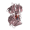
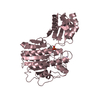
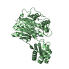
- PDB-6pld: Crystal Structure of Pseudomonas aeruginosa D-Arginine Dehydrogen... -
+ Open data

- Basic information
| Entry | Database: PDB / ID: 6pld | ||||||
|---|---|---|---|---|---|---|---|
| Title | Crystal Structure of Pseudomonas aeruginosa D-Arginine Dehydrogenase Y249F variant with 6-OH-FAD - Green fraction | ||||||
 Components Components | FAD-dependent catabolic D-arginine dehydrogenase DauA | ||||||
 Keywords Keywords | FLAVOPROTEIN / Flavin / 6-OH-FAD | ||||||
| Function / homology |  Function and homology information Function and homology informationD-arginine dehydrogenase / D-amino-acid dehydrogenase activity / arginine metabolic process / L-arginine catabolic process / nucleotide binding / cytoplasm Similarity search - Function | ||||||
| Biological species |   Pseudomonas aeruginosa (bacteria) Pseudomonas aeruginosa (bacteria) | ||||||
| Method |  X-RAY DIFFRACTION / SYNCHROTRON / MOLECULAR REPLACEMENT / Resolution: 1.55 Å X-RAY DIFFRACTION / SYNCHROTRON / MOLECULAR REPLACEMENT / Resolution: 1.55 Å | ||||||
 Authors Authors | Reis, R.A.G. / Iyer, A. / Agniswamy, J. / Gannavaram, S. / Weber, I. / Gadda, G. | ||||||
| Funding support |  United States, 1items United States, 1items
| ||||||
 Citation Citation | Journal: Biochemistry / Year: 2021 Title: A Single-Point Mutation in d-Arginine Dehydrogenase Unlocks a Transient Conformational State Resulting in Altered Cofactor Reactivity. Authors: Iyer, A. / Reis, R.A.G. / Gannavaram, S. / Momin, M. / Spring-Connell, A.M. / Orozco-Gonzalez, Y. / Agniswamy, J. / Hamelberg, D. / Weber, I.T. / Gozem, S. / Wang, S. / Germann, M.W. / Gadda, G. | ||||||
| History |
|
- Structure visualization
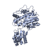
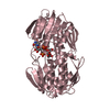
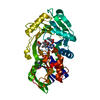
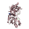
| Structure viewer | Molecule: MolmilJmol/JSmol |
|---|
- Downloads & links
-Download
| PDBx/mmCIF format | 6pld.cif.gz | 106.4 KB | Display | PDBx/mmCIF format |
|---|---|---|---|---|
| PDB format | pdb6pld.ent.gz | 77.4 KB | Display | PDB format |
| PDBx/mmJSON format | 6pld.json.gz | Tree view | PDBx/mmJSON format | |
| Others |  Other downloads Other downloads |
-Validation report
| Arichive directory | https://data.pdbj.org/pub/pdb/validation_reports/pl/6pldftp://data.pdbj.org/pub/pdb/validation_reports/pl/6pld | HTTPS FTP |
|---|
-Related structure data
| Related structure data |  6p9dC 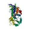 3nyeS C: citing same article ( S: Starting model for refinement |
|---|---|
| Similar structure data |


-Links
 PDBj
PDBj- Assembly
| Deposited unit | 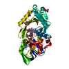
| ||||||||
|---|---|---|---|---|---|---|---|---|---|
| 1 |
| ||||||||
| Unit cell |
|
-Components
| #1: Protein | Mass: 40594.715 Da / Num. of mol.: 1 / Mutation: Y249F Source method: isolated from a genetically manipulated source Source: (gene. exp.) Pseudomonas aeruginosa (strain ATCC 15692 / DSM 22644 / CIP 104116 / JCM 14847 / LMG 12228 / 1C / PRS 101 / PAO1) (bacteria)Strain: ATCC 15692 / DSM 22644 / CIP 104116 / JCM 14847 / LMG 12228 / 1C / PRS 101 / PAO1 Gene: dauA, PA3863 / Production host: | ||||
|---|---|---|---|---|---|
| #2: Chemical | ChemComp-6FA / 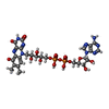  Mass: 801.549 Da / Num. of mol.: 1 / Source method: obtained synthetically / Formula: C27H33N9O16P2 / Feature type: SUBJECT OF INVESTIGATION Mass: 801.549 Da / Num. of mol.: 1 / Source method: obtained synthetically / Formula: C27H33N9O16P2 / Feature type: SUBJECT OF INVESTIGATION | ||||
| #3: Chemical | ChemComp-PEG /   Mass: 106.120 Da / Num. of mol.: 1 / Source method: obtained synthetically / Formula: C4H10O3 Mass: 106.120 Da / Num. of mol.: 1 / Source method: obtained synthetically / Formula: C4H10O3 | ||||
| #4: Chemical | ChemComp-GOL /   Mass: 92.094 Da / Num. of mol.: 6 / Source method: obtained synthetically / Formula: C3H8O3 Mass: 92.094 Da / Num. of mol.: 6 / Source method: obtained synthetically / Formula: C3H8O3#5: Water | ChemComp-HOH / |  Mass: 18.015 Da / Num. of mol.: 364 / Source method: isolated from a natural source / Formula: H2O Mass: 18.015 Da / Num. of mol.: 364 / Source method: isolated from a natural source / Formula: H2OHas ligand of interest | Y | |
-Experimental details
-Experiment
| Experiment | Method: X-RAY DIFFRACTION / Number of used crystals: 1 |
|---|
- Sample preparation
| Crystal | Density Matthews: 2.09 Å3/Da / Density % sol: 39.01 % |
|---|---|
| Crystal grow | Temperature: 298 K / Method: vapor diffusion, hanging drop / pH: 6.8 Details: 0.05 M Tris-Cl pH 6.5-7.0, 10% glycerol, and 12-13% (w/v) PEG 3350, 5-10% PEG 5000 PH range: 6.5-7.0 |
-Data collection
| Diffraction | Mean temperature: 100 K / Serial crystal experiment: N |
|---|---|
| Diffraction source | Source: SYNCHROTRON / Site: APS / Beamline: 22-ID / Wavelength: 1 Å |
| Detector | Type: DECTRIS PILATUS 2M / Detector: PIXEL / Date: Feb 11, 2017 |
| Radiation | Protocol: SINGLE WAVELENGTH / Monochromatic (M) / Laue (L): M / Scattering type: x-ray |
| Radiation wavelength | Wavelength: 1 Å / Relative weight: 1 |
| Reflection | Resolution: 1.55→59.78 Å / Num. obs: 48055 / % possible obs: 96.1 % / Redundancy: 7 % / Biso Wilson estimate: 12.484 Å2 / CC1/2: 0.995 / Rmerge(I) obs: 0.103 / Net I/σ(I): 10.6 |
| Reflection shell | Resolution: 1.55→1.58 Å / Redundancy: 6.5 % / Rmerge(I) obs: 0.488 / Mean I/σ(I) obs: 3.2 / Num. unique obs: 2284 / CC1/2: 0.896 / % possible all: 93.4 |
- Processing
| Software |
| ||||||||||||||||||||||||||||||||||||||||||||||||||||||||||||||||||||||||||||||||||||||||||||||||||||||||||||||||||||||||||||||
|---|---|---|---|---|---|---|---|---|---|---|---|---|---|---|---|---|---|---|---|---|---|---|---|---|---|---|---|---|---|---|---|---|---|---|---|---|---|---|---|---|---|---|---|---|---|---|---|---|---|---|---|---|---|---|---|---|---|---|---|---|---|---|---|---|---|---|---|---|---|---|---|---|---|---|---|---|---|---|---|---|---|---|---|---|---|---|---|---|---|---|---|---|---|---|---|---|---|---|---|---|---|---|---|---|---|---|---|---|---|---|---|---|---|---|---|---|---|---|---|---|---|---|---|---|---|---|---|
| Refinement | Method to determine structure: MOLECULAR REPLACEMENT Starting model: 3NYE Resolution: 1.55→39.789 Å / SU ML: 0.16 / Cross valid method: FREE R-VALUE / σ(F): 1.34 / Phase error: 18.58
| ||||||||||||||||||||||||||||||||||||||||||||||||||||||||||||||||||||||||||||||||||||||||||||||||||||||||||||||||||||||||||||||
| Solvent computation | Shrinkage radii: 0.9 Å / VDW probe radii: 1.11 Å | ||||||||||||||||||||||||||||||||||||||||||||||||||||||||||||||||||||||||||||||||||||||||||||||||||||||||||||||||||||||||||||||
| Refinement step | Cycle: LAST / Resolution: 1.55→39.789 Å
| ||||||||||||||||||||||||||||||||||||||||||||||||||||||||||||||||||||||||||||||||||||||||||||||||||||||||||||||||||||||||||||||
| Refine LS restraints |
| ||||||||||||||||||||||||||||||||||||||||||||||||||||||||||||||||||||||||||||||||||||||||||||||||||||||||||||||||||||||||||||||
| LS refinement shell |
|
