 Movie
Movie Controller

[English] 日本語
 Yorodumi
- PDB-6pe3: Structure of YcaO enzyme from Methanocaldococcus jannaschii in co... -
+ Open data

- Basic information
| Entry | Database: PDB / ID: 6pe3 | ||||||
|---|---|---|---|---|---|---|---|
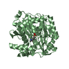
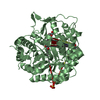
| Title | Structure of YcaO enzyme from Methanocaldococcus jannaschii in complex with ATP | ||||||
 Components Components | Uncharacterized protein MJ1094 | ||||||
 Keywords Keywords | BIOSYNTHETIC PROTEIN / YcaO / ATP / thioamide / Methanocaldococcus jannaschii | ||||||
| Function / homology | Putative methanogenesis marker protein 1 / YcaO-like domain / YcaO cyclodehydratase, ATP-ad Mg2+-binding / YcaO domain profile. / cytosol / ADENOSINE-5'-TRIPHOSPHATE / Uncharacterized protein MJ1094 Function and homology information Function and homology information | ||||||
| Biological species |   Methanocaldococcus jannaschii (archaea) Methanocaldococcus jannaschii (archaea) | ||||||
| Method |  X-RAY DIFFRACTION / SYNCHROTRON / MOLECULAR REPLACEMENT / Resolution: 2.3 Å X-RAY DIFFRACTION / SYNCHROTRON / MOLECULAR REPLACEMENT / Resolution: 2.3 Å | ||||||
 Authors Authors | Dong, S.-H. / Nair, S.K. | ||||||
| Funding support |  United States, 1items United States, 1items
| ||||||
 Citation Citation | Journal: Acs Cent.Sci. / Year: 2019 Title: Mechanistic Basis for Ribosomal Peptide Backbone Modifications. Authors: Dong, S.H. / Liu, A. / Mahanta, N. / Mitchell, D.A. / Nair, S.K. | ||||||
| History |
|
- Structure visualization
| Structure viewer | Molecule: MolmilJmol/JSmol |
|---|
- Downloads & links
-Download
| PDBx/mmCIF format | 6pe3.cif.gz | 94.1 KB | Display | PDBx/mmCIF format |
|---|---|---|---|---|
| PDB format | pdb6pe3.ent.gz | 69.9 KB | Display | PDB format |
| PDBx/mmJSON format | 6pe3.json.gz | Tree view | PDBx/mmJSON format | |
| Others |  Other downloads Other downloads |
-Validation report
| Arichive directory | https://data.pdbj.org/pub/pdb/validation_reports/pe/6pe3ftp://data.pdbj.org/pub/pdb/validation_reports/pe/6pe3 | HTTPS FTP |
|---|
-Related structure data


-Links
 PDBj
PDBj- Assembly
| Deposited unit | 
| |||||||||
|---|---|---|---|---|---|---|---|---|---|---|
| 1 |
| |||||||||
| Unit cell |
| |||||||||
| Components on special symmetry positions |
|
-Components
| #1: Protein | Mass: 44228.992 Da / Num. of mol.: 1 Source method: isolated from a genetically manipulated source Source: (gene. exp.) Methanocaldococcus jannaschii (strain ATCC 43067 / DSM 2661 / JAL-1 / JCM 10045 / NBRC 100440) (archaea)Strain: ATCC 43067 / DSM 2661 / JAL-1 / JCM 10045 / NBRC 100440 Gene: MJ1094 / Production host:  |
|---|---|
| #2: Chemical | ChemComp-ATP /   Mass: 507.181 Da / Num. of mol.: 1 / Source method: obtained synthetically / Formula: C10H16N5O13P3 / Feature type: SUBJECT OF INVESTIGATION / Comment: ATP, energy-carrying molecule*YM Mass: 507.181 Da / Num. of mol.: 1 / Source method: obtained synthetically / Formula: C10H16N5O13P3 / Feature type: SUBJECT OF INVESTIGATION / Comment: ATP, energy-carrying molecule*YM |
| #3: Water | ChemComp-HOH /  Mass: 18.015 Da / Num. of mol.: 172 / Source method: isolated from a natural source / Formula: H2O Mass: 18.015 Da / Num. of mol.: 172 / Source method: isolated from a natural source / Formula: H2O |
| Has ligand of interest | Y |
-Experimental details
-Experiment
| Experiment | Method: X-RAY DIFFRACTION / Number of used crystals: 1 |
|---|
- Sample preparation
| Crystal | Density Matthews: 3.53 Å3/Da / Density % sol: 65.17 % |
|---|---|
| Crystal grow | Temperature: 282 K / Method: vapor diffusion, hanging drop Details: 0.1 M sodium acetate pH 4.6, 2.0 M ammonium sulfate, 0.02 M taurine |
-Data collection
| Diffraction | Mean temperature: 80 K / Ambient temp details: liquid nitrogen flow / Serial crystal experiment: N |
|---|---|
| Diffraction source | Source: SYNCHROTRON / Site: APS / Beamline: 21-ID-D / Wavelength: 0.979106 Å |
| Detector | Type: DECTRIS EIGER X 9M / Detector: PIXEL / Date: Aug 5, 2017 |
| Radiation | Protocol: SINGLE WAVELENGTH / Monochromatic (M) / Laue (L): M / Scattering type: x-ray |
| Radiation wavelength | Wavelength: 0.979106 Å / Relative weight: 1 |
| Reflection | Resolution: 2.3→25.6 Å / Num. obs: 26776 / % possible obs: 95.61 % / Redundancy: 30.1 % / Net I/σ(I): 19.9 |
| Reflection shell | Resolution: 2.3→2.34 Å / Num. unique obs: 1846 |
- Processing
| Software |
| ||||||||||||||||||||||||||||||||||||||||||||||||||||||||||||||||||||||||||||||||||||||||||||||||||||||||||||||||||||||||||||||||||||||||||||||||||||||||||||||||||||||||||||||||||||||
|---|---|---|---|---|---|---|---|---|---|---|---|---|---|---|---|---|---|---|---|---|---|---|---|---|---|---|---|---|---|---|---|---|---|---|---|---|---|---|---|---|---|---|---|---|---|---|---|---|---|---|---|---|---|---|---|---|---|---|---|---|---|---|---|---|---|---|---|---|---|---|---|---|---|---|---|---|---|---|---|---|---|---|---|---|---|---|---|---|---|---|---|---|---|---|---|---|---|---|---|---|---|---|---|---|---|---|---|---|---|---|---|---|---|---|---|---|---|---|---|---|---|---|---|---|---|---|---|---|---|---|---|---|---|---|---|---|---|---|---|---|---|---|---|---|---|---|---|---|---|---|---|---|---|---|---|---|---|---|---|---|---|---|---|---|---|---|---|---|---|---|---|---|---|---|---|---|---|---|---|---|---|---|---|
| Refinement | Method to determine structure: MOLECULAR REPLACEMENT / Resolution: 2.3→25 Å / Cor.coef. Fo:Fc: 0.963 / Cor.coef. Fo:Fc free: 0.935 / SU B: 8.194 / SU ML: 0.183 / Cross valid method: THROUGHOUT / ESU R: 0.241 / ESU R Free: 0.212 / Details: HYDROGENS HAVE BEEN USED IF PRESENT IN THE INPUT
| ||||||||||||||||||||||||||||||||||||||||||||||||||||||||||||||||||||||||||||||||||||||||||||||||||||||||||||||||||||||||||||||||||||||||||||||||||||||||||||||||||||||||||||||||||||||
| Solvent computation | Ion probe radii: 0.8 Å / Shrinkage radii: 0.8 Å / VDW probe radii: 1.2 Å | ||||||||||||||||||||||||||||||||||||||||||||||||||||||||||||||||||||||||||||||||||||||||||||||||||||||||||||||||||||||||||||||||||||||||||||||||||||||||||||||||||||||||||||||||||||||
| Displacement parameters | Biso mean: 68.259 Å2
| ||||||||||||||||||||||||||||||||||||||||||||||||||||||||||||||||||||||||||||||||||||||||||||||||||||||||||||||||||||||||||||||||||||||||||||||||||||||||||||||||||||||||||||||||||||||
| Refinement step | Cycle: 1 / Resolution: 2.3→25 Å
| ||||||||||||||||||||||||||||||||||||||||||||||||||||||||||||||||||||||||||||||||||||||||||||||||||||||||||||||||||||||||||||||||||||||||||||||||||||||||||||||||||||||||||||||||||||||
| Refine LS restraints |
|