 Movie
Movie Controller

[English] 日本語
 Yorodumi
- PDB-6oyc: Glycosylation Associate Protein (Gap123) complex from Streptococc... -
+ Open data

- Basic information
| Entry | Database: PDB / ID: 6oyc | ||||||
|---|---|---|---|---|---|---|---|
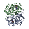
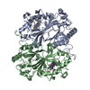
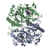
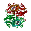
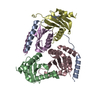
| Title | Glycosylation Associate Protein (Gap123) complex from Streptococcus agalactiae | ||||||
 Components Components | (Glycosylation Associate Protein ...) x 3 | ||||||
 Keywords Keywords | SUGAR BINDING PROTEIN / Glycosylation / Secretion / lectin | ||||||
| Function / homology |  Function and homology information Function and homology information | ||||||
| Biological species | Streptococcus agalactiae serotype V | ||||||
| Method |  X-RAY DIFFRACTION / SYNCHROTRON / MOLECULAR REPLACEMENT / Resolution: 2.351 Å X-RAY DIFFRACTION / SYNCHROTRON / MOLECULAR REPLACEMENT / Resolution: 2.351 Å | ||||||
 Authors Authors | zhang, H. / Wu, H. | ||||||
| Funding support |  United States, 1items United States, 1items
| ||||||
 Citation Citation | Journal: To Be Published Title: Glycosylation Associate Protein (Gap123) complex from Streptococcus agalactiae Authors: zhang, H. / Zhu, F. / Wu, H. | ||||||
| History |
|
- Structure visualization
| Structure viewer | Molecule: MolmilJmol/JSmol |
|---|
- Downloads & links
-Download
| PDBx/mmCIF format | 6oyc.cif.gz | 289.7 KB | Display | PDBx/mmCIF format |
|---|---|---|---|---|
| PDB format | pdb6oyc.ent.gz | 230.4 KB | Display | PDB format |
| PDBx/mmJSON format | 6oyc.json.gz | Tree view | PDBx/mmJSON format | |
| Others |  Other downloads Other downloads |
-Validation report
| Summary document | 6oyc_validation.pdf.gz | 280.5 KB | Display | wwPDB validaton report |
|---|---|---|---|---|
| Full document | 6oyc_full_validation.pdf.gz | 281.8 KB | Display | |
| Data in XML | 6oyc_validation.xml.gz | 2 KB | Display | |
| Data in CIF | 6oyc_validation.cif.gz | 16.6 KB | Display | |
| Arichive directory | https://data.pdbj.org/pub/pdb/validation_reports/oy/6oycftp://data.pdbj.org/pub/pdb/validation_reports/oy/6oyc | HTTPS FTP |
-Related structure data
| Related structure data |  6e2i S: Starting model for refinement |
|---|---|
| Similar structure data |

-Links
 PDBj
PDBj- Assembly
| Deposited unit | 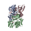
| ||||||||
|---|---|---|---|---|---|---|---|---|---|
| 1 | 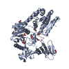
| ||||||||
| 2 | 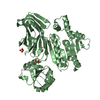
| ||||||||
| 3 | 
| ||||||||
| 4 |
| ||||||||
| Unit cell |
| ||||||||
| Components on special symmetry positions |
|
-Components
-Glycosylation Associate Protein ... , 3 types, 3 molecules ABC
| #1: Protein | Mass: 60541.078 Da / Num. of mol.: 1 Source method: isolated from a genetically manipulated source Source: (gene. exp.)  Streptococcus agalactiae serotype V (strain ATCC BAA-611 / 2603 V/R) (bacteria) Streptococcus agalactiae serotype V (strain ATCC BAA-611 / 2603 V/R) (bacteria)Strain: ATCC BAA-611 / 2603 V/R / Gene: SAG1452 / Production host: |
|---|---|
| #2: Protein | Mass: 59251.688 Da / Num. of mol.: 1 Source method: isolated from a genetically manipulated source Source: (gene. exp.) Streptococcus agalactiae serotype V (strain ATCC BAA-611 / 2603 V/R) (bacteria)Strain: ATCC BAA-611 / 2603 V/R / Gene: SAG1451 / Production host: |
| #3: Protein | Mass: 37928.027 Da / Num. of mol.: 1 Source method: isolated from a genetically manipulated source Source: (gene. exp.) Streptococcus agalactiae serotype V (strain ATCC BAA-611 / 2603 V/R) (bacteria)Strain: ATCC BAA-611 / 2603 V/R / Gene: SAG1450 / Production host: |
-Non-polymers , 3 types, 419 molecules 


| #4: Chemical | ChemComp-GOL /  Mass: 92.094 Da / Num. of mol.: 13 / Source method: obtained synthetically / Formula: C3H8O3 Mass: 92.094 Da / Num. of mol.: 13 / Source method: obtained synthetically / Formula: C3H8O3#5: Chemical |  Mass: 96.063 Da / Num. of mol.: 3 / Source method: obtained synthetically / Formula: SO4 Mass: 96.063 Da / Num. of mol.: 3 / Source method: obtained synthetically / Formula: SO4#6: Water | ChemComp-HOH / | Mass: 18.015 Da / Num. of mol.: 403 / Source method: isolated from a natural source / Formula: H2O |
|---|
-Experimental details
-Experiment
| Experiment | Method: X-RAY DIFFRACTION / Number of used crystals: 1 |
|---|
- Sample preparation
| Crystal | Density Matthews: 2.83 Å3/Da / Density % sol: 56.54 % |
|---|---|
| Crystal grow | Temperature: 293 K / Method: vapor diffusion, sitting drop / Details: 0.1M HEPES pH 7.5, 5% PEG 400, 2M Ammonium sulfate |
-Data collection
| Diffraction | Mean temperature: 100 K / Serial crystal experiment: N |
|---|---|
| Diffraction source | Source: SYNCHROTRON / Site: APS / Beamline: 22-ID / Wavelength: 1 Å |
| Detector | Type: DECTRIS EIGER X 16M / Detector: PIXEL / Date: Feb 15, 2017 |
| Radiation | Protocol: SINGLE WAVELENGTH / Monochromatic (M) / Laue (L): M / Scattering type: x-ray |
| Radiation wavelength | Wavelength: 1 Å / Relative weight: 1 |
| Reflection | Resolution: 2.35→49.272 Å / Num. obs: 72701 / % possible obs: 99.55 % / Redundancy: 14.1 % / Net I/σ(I): 28.13 |
| Reflection shell | Resolution: 2.35→2.43 Å / Num. unique obs: 7084 |
- Processing
| Software |
| |||||||||||||||||||||||||||||||||||||||||||||||||||||||||||||||||||||||||||||||||||||||||||||||||||||||||
|---|---|---|---|---|---|---|---|---|---|---|---|---|---|---|---|---|---|---|---|---|---|---|---|---|---|---|---|---|---|---|---|---|---|---|---|---|---|---|---|---|---|---|---|---|---|---|---|---|---|---|---|---|---|---|---|---|---|---|---|---|---|---|---|---|---|---|---|---|---|---|---|---|---|---|---|---|---|---|---|---|---|---|---|---|---|---|---|---|---|---|---|---|---|---|---|---|---|---|---|---|---|---|---|---|---|---|
| Refinement | Method to determine structure: MOLECULAR REPLACEMENT Starting model: 6E2I 6e2i Resolution: 2.351→49.272 Å / SU ML: 0.27 / Cross valid method: FREE R-VALUE / σ(F): 1.36 / Phase error: 23.21
| |||||||||||||||||||||||||||||||||||||||||||||||||||||||||||||||||||||||||||||||||||||||||||||||||||||||||
| Solvent computation | Shrinkage radii: 0.9 Å / VDW probe radii: 1.11 Å | |||||||||||||||||||||||||||||||||||||||||||||||||||||||||||||||||||||||||||||||||||||||||||||||||||||||||
| Refinement step | Cycle: LAST / Resolution: 2.351→49.272 Å
| |||||||||||||||||||||||||||||||||||||||||||||||||||||||||||||||||||||||||||||||||||||||||||||||||||||||||
| Refine LS restraints |
| |||||||||||||||||||||||||||||||||||||||||||||||||||||||||||||||||||||||||||||||||||||||||||||||||||||||||
| LS refinement shell |
|
