- PDB-6ovi: Crystal Structure of KDPG Aldolase from Legionella Pneumophila wi... -
+
Open data
ID or keywords:
Loading...
-
Basic information
Entry
Database: PDB / ID: 6ovi
Title
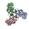
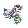
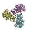
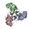
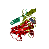
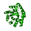
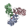
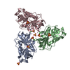
Crystal Structure of KDPG Aldolase from Legionella Pneumophila with pyruvate captured at low pH as a covalent carbinolamine intermediate
Components
Keto-deoxy-phosphogluconate aldolase
Keywords
LYASE / SSGCID / KDPG ALDOLASE / Structural Genomics / Seattle Structural Genomics Center for Infectious Disease
Function / homology
Function and homology information
2-dehydro-3-deoxy-phosphogluconate aldolase / 2-dehydro-3-deoxy-phosphogluconate aldolase activity Similarity search - Function
KDPG/KHG aldolase, active site 2 / KDPG and KHG aldolases Schiff-base forming residue. / KDPG/KHG aldolase, active site 1 / KDPG and KHG aldolases active site. / KDPG/KHG aldolase / KDPG and KHG aldolase / Aldolase class I / Aldolase-type TIM barrel / TIM Barrel / Alpha-Beta Barrel / Alpha Beta Similarity search - Domain/homology
National Institutes of Health/National Institute Of Allergy and Infectious Diseases (NIH/NIAID)
United States
Citation
Journal: To Be Published Title: Crystal Structure of KDPG Aldolase from Legionella Pneumophila with pyruvate captured at low pH as a covalent carbinolamine intermediate Authors: Davies, D.R. / Dranow, D.M.
In the structure databanks used in Yorodumi, some data are registered as the other names, "COVID-19 virus" and "2019-nCoV". Here are the details of the virus and the list of structure data.
Jan 31, 2019. EMDB accession codes are about to change! (news from PDBe EMDB page)
EMDB accession codes are about to change! (news from PDBe EMDB page)
The allocation of 4 digits for EMDB accession codes will soon come to an end. Whilst these codes will remain in use, new EMDB accession codes will include an additional digit and will expand incrementally as the available range of codes is exhausted. The current 4-digit format prefixed with “EMD-” (i.e. EMD-XXXX) will advance to a 5-digit format (i.e. EMD-XXXXX), and so on. It is currently estimated that the 4-digit codes will be depleted around Spring 2019, at which point the 5-digit format will come into force.
The EM Navigator/Yorodumi systems omit the EMD- prefix.
Related info.:Q: What is EMD? / ID/Accession-code notation in Yorodumi/EM Navigator
Yorodumi is a browser for structure data from EMDB, PDB, SASBDB, etc.
This page is also the successor to EM Navigator detail page, and also detail information page/front-end page for Omokage search.
The word "yorodu" (or yorozu) is an old Japanese word meaning "ten thousand". "mi" (miru) is to see.
Related info.:EMDB / PDB / SASBDB / Comparison of 3 databanks / Yorodumi Search / Aug 31, 2016. New EM Navigator & Yorodumi / Yorodumi Papers / Jmol/JSmol / Function and homology information / Changes in new EM Navigator and Yorodumi
 Movie
Movie Controller

 Yorodumi
 Open data
 Basic information
 Components
 Keywords
 Function and homology information
 Legionella pneumophila (bacteria)
 X-RAY DIFFRACTION /
  Authors
 United States, 1items
  Citation
 Structure visualization
 Downloads & links
 Other downloads


 PDBj
PDBj Assembly
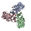
 Type: L-peptide linking / Mass: 106.077 Da / Num. of mol.: 3 / Source method: obtained synthetically / Formula: C3H6O4 / Feature type: SUBJECT OF INVESTIGATION
 Mass: 62.068 Da / Num. of mol.: 4 / Source method: obtained synthetically / Formula: C2H6O2
 Mass: 96.063 Da / Num. of mol.: 6 / Source method: obtained synthetically / Formula: SO4
 Mass: 18.015 Da / Num. of mol.: 524 / Source method: isolated from a natural source / Formula: H2O
 Sample preparation
 Processing