 Movie
Movie Controller

[English] 日本語
 Yorodumi
- PDB-6ous: Structure of fusion glycoprotein from human respiratory syncytial... -
+ Open data

- Basic information
| Entry | Database: PDB / ID: 6ous | ||||||||||||
|---|---|---|---|---|---|---|---|---|---|---|---|---|---|
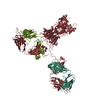
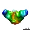
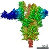
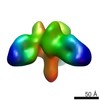
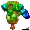
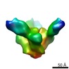
| Title | Structure of fusion glycoprotein from human respiratory syncytial virus | ||||||||||||
 Components Components |
| ||||||||||||
 Keywords Keywords | VIRAL PROTEIN/Immune system / Antibody / RSV / Neutralizing / VIRAL PROTEIN / VIRAL PROTEIN-Immune system complex | ||||||||||||
| Function / homology |  Function and homology information Function and homology informationsymbiont-mediated induction of syncytium formation / Translation of respiratory syncytial virus mRNAs / RSV-host interactions / Assembly and release of respiratory syncytial virus (RSV) virions / Maturation of hRSV A proteins / Respiratory syncytial virus (RSV) attachment and entry / host cell Golgi membrane / entry receptor-mediated virion attachment to host cell / fusion of virus membrane with host plasma membrane / viral envelope ...symbiont-mediated induction of syncytium formation / Translation of respiratory syncytial virus mRNAs / RSV-host interactions / Assembly and release of respiratory syncytial virus (RSV) virions / Maturation of hRSV A proteins / Respiratory syncytial virus (RSV) attachment and entry / host cell Golgi membrane / entry receptor-mediated virion attachment to host cell / fusion of virus membrane with host plasma membrane / viral envelope / symbiont entry into host cell / host cell plasma membrane / virion membrane / identical protein binding / plasma membrane Similarity search - Function | ||||||||||||
| Biological species |  Human respiratory syncytial virus A2 Human respiratory syncytial virus A2 Human immunodeficiency virus 1 Human immunodeficiency virus 1 Homo sapiens (human) Homo sapiens (human) | ||||||||||||
| Method |  X-RAY DIFFRACTION / SYNCHROTRON / MOLECULAR REPLACEMENT / molecular replacement / Resolution: 3.4 Å X-RAY DIFFRACTION / SYNCHROTRON / MOLECULAR REPLACEMENT / molecular replacement / Resolution: 3.4 Å | ||||||||||||
 Authors Authors | Su, H.P. | ||||||||||||
 Citation Citation | Journal: Nat Commun / Year: 2019 Title: A potent broadly neutralizing human RSV antibody targets conserved site IV of the fusion glycoprotein. Authors: Tang, A. / Chen, Z. / Cox, K.S. / Su, H.P. / Callahan, C. / Fridman, A. / Zhang, L. / Patel, S.B. / Cejas, P.J. / Swoyer, R. / Touch, S. / Citron, M.P. / Govindarajan, D. / Luo, B. / Eddins, ...Authors: Tang, A. / Chen, Z. / Cox, K.S. / Su, H.P. / Callahan, C. / Fridman, A. / Zhang, L. / Patel, S.B. / Cejas, P.J. / Swoyer, R. / Touch, S. / Citron, M.P. / Govindarajan, D. / Luo, B. / Eddins, M. / Reid, J.C. / Soisson, S.M. / Galli, J. / Wang, D. / Wen, Z. / Heidecker, G.J. / Casimiro, D.R. / DiStefano, D.J. / Vora, K.A. | ||||||||||||
| History |
|
- Structure visualization
| Structure viewer | Molecule: MolmilJmol/JSmol |
|---|
- Downloads & links
-Download
| PDBx/mmCIF format | 6ous.cif.gz | 1.9 MB | Display | PDBx/mmCIF format |
|---|---|---|---|---|
| PDB format | pdb6ous.ent.gz | 1.6 MB | Display | PDB format |
| PDBx/mmJSON format | 6ous.json.gz | Tree view | PDBx/mmJSON format | |
| Others |  Other downloads Other downloads |
-Validation report
| Arichive directory | https://data.pdbj.org/pub/pdb/validation_reports/ou/6ousftp://data.pdbj.org/pub/pdb/validation_reports/ou/6ous | HTTPS FTP |
|---|
-Related structure data


-Links
 PDBj
PDBj


- Assembly
| Deposited unit | 
| ||||||||||
|---|---|---|---|---|---|---|---|---|---|---|---|
| 1 | 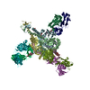
| ||||||||||
| 2 | 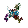
| ||||||||||
| Unit cell |
|
-Components
-Fusion glycoprotein ... , 2 types, 12 molecules ACEGIKBDFHJL
| #1: Protein | Mass: 9512.768 Da / Num. of mol.: 6 Source method: isolated from a genetically manipulated source Source: (gene. exp.) Human respiratory syncytial virus A2 / Strain: A2 / Production host: Homo sapiens (human) / References: UniProt: P03420#2: Protein | Mass: 45609.148 Da / Num. of mol.: 6 Source method: isolated from a genetically manipulated source Source: (gene. exp.) Human respiratory syncytial virus A2, (gene. exp.) Human immunodeficiency virus 1Strain: A2 / Production host: Homo sapiens (human) / References: UniProt: P03420, UniProt: M1E1E4 |
|---|
-Antibody , 2 types, 12 molecules MOQSUWNPRTVX
| #3: Antibody | Mass: 24551.439 Da / Num. of mol.: 6 Source method: isolated from a genetically manipulated source Source: (gene. exp.) Homo sapiens (human) / Production host:   Cricetulus griseus (Chinese hamster) Cricetulus griseus (Chinese hamster)#4: Antibody | Mass: 23406.043 Da / Num. of mol.: 6 Source method: isolated from a genetically manipulated source Source: (gene. exp.) Homo sapiens (human) / Production host: Cricetulus griseus (Chinese hamster) |
|---|
-Sugars , 2 types, 10 molecules 
| #5: Polysaccharide | alpha-D-mannopyranose-(1-3)-beta-D-mannopyranose-(1-4)-2-acetamido-2-deoxy-beta-D-glucopyranose-(1- ...alpha-D-mannopyranose-(1-3)-beta-D-mannopyranose-(1-4)-2-acetamido-2-deoxy-beta-D-glucopyranose-(1-4)-2-acetamido-2-deoxy-beta-D-glucopyranose Source method: isolated from a genetically manipulated source #6: Sugar | ChemComp-NAG /  Type: D-saccharide, beta linking / Mass: 221.208 Da / Num. of mol.: 4 Type: D-saccharide, beta linking / Mass: 221.208 Da / Num. of mol.: 4Source method: isolated from a genetically manipulated source Formula: C8H15NO6 |
|---|
-Details
| Has ligand of interest | N |
|---|---|
| Has protein modification | Y |
-Experimental details
-Experiment
| Experiment | Method: X-RAY DIFFRACTION / Number of used crystals: 1 |
|---|
- Sample preparation
| Crystal | Density Matthews: 3.37 Å3/Da / Density % sol: 63.51 % |
|---|---|
| Crystal grow | Temperature: 293 K / Method: vapor diffusion, hanging drop / pH: 7.5 Details: 100 mM Tris 8.5, 10% PEG 8000, 200 mM ammonium sulfate |
-Data collection
| Diffraction | Mean temperature: 100 K / Serial crystal experiment: N | |||||||||||||||||||||||||||
|---|---|---|---|---|---|---|---|---|---|---|---|---|---|---|---|---|---|---|---|---|---|---|---|---|---|---|---|---|
| Diffraction source | Source: SYNCHROTRON / Site: CLSI  / Beamline: 08ID-1 / Wavelength: 0.97949 Å / Beamline: 08ID-1 / Wavelength: 0.97949 Å | |||||||||||||||||||||||||||
| Detector | Type: RAYONIX MX300HE / Detector: CCD / Date: May 17, 2015 | |||||||||||||||||||||||||||
| Radiation | Protocol: SINGLE WAVELENGTH / Monochromatic (M) / Laue (L): M / Scattering type: x-ray | |||||||||||||||||||||||||||
| Radiation wavelength | Wavelength: 0.97949 Å / Relative weight: 1 | |||||||||||||||||||||||||||
| Reflection | Resolution: 3.4→49.86 Å / Num. obs: 114498 / % possible obs: 99 % / Redundancy: 2 % / Biso Wilson estimate: 78.54 Å2 / CC1/2: 0.98 / Rmerge(I) obs: 0.127 / Rpim(I) all: 0.127 / Rrim(I) all: 0.179 / Net I/σ(I): 3.9 / Num. measured all: 228565 | |||||||||||||||||||||||||||
| Reflection shell | Diffraction-ID: 1 / Redundancy: 2 %
|
-Phasing
| Phasing | Method: molecular replacement | |||||||||
|---|---|---|---|---|---|---|---|---|---|---|
| Phasing MR | Model details: Phaser MODE: MR_AUTO
|
- Processing
| Software |
| ||||||||||||||||||
|---|---|---|---|---|---|---|---|---|---|---|---|---|---|---|---|---|---|---|---|
| Refinement | Method to determine structure: MOLECULAR REPLACEMENT Starting model: 4mmt, 1hzh Resolution: 3.4→49.86 Å / Cross valid method: FREE R-VALUE
| ||||||||||||||||||
| Refinement step | Cycle: LAST / Resolution: 3.4→49.86 Å
|
