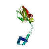
 Movie
Movie Controller


+ Open data

- Basic information
| Entry | Database: PDB / ID: 6oqe | ||||||
|---|---|---|---|---|---|---|---|
| Title | X-ray structure of H6N6-NS1 delta(80-84) R38A K41A mutant | ||||||
 Components Components | Non-structural protein 1 | ||||||
 Keywords Keywords |  VIRAL PROTEIN / Protein VIRAL PROTEIN / Protein | ||||||
| Function / homology |  Function and homology information Function and homology informationsymbiont-mediated suppression of host mRNA processing / symbiont-mediated suppression of host PKR/eIFalpha signaling / protein serine/threonine kinase inhibitor activity / symbiont-mediated suppression of host cytoplasmic pattern recognition receptor signaling pathway via inhibition of RIG-I activity / host cell cytoplasm / symbiont-mediated suppression of host type I interferon-mediated signaling pathway / host cell nucleus / RNA bindingSimilarity search - Function | ||||||
| Biological species |   Influenza A virus Influenza A virus | ||||||
| Method | X-RAY DIFFRACTION / SYNCHROTRON / MOLECULAR REPLACEMENT / Resolution: 3.899 Å | ||||||
 Authors Authors | Mitra, S. / Kumar, D. / Hu, L. / Prasad, B.V.V. | ||||||
| Funding support |  United States, 1items United States, 1items
| ||||||
 Citation Citation | Journal: J.Virol. / Year: 2019 Title: Influenza A Virus Protein NS1 Exhibits Strain-Independent Conformational Plasticity. Authors: Mitra, S. / Kumar, D. / Hu, L. / Sankaran, B. / Moosa, M.M. / Rice, A.P. / Ferreon, J.C. / Ferreon, A.C.M. / Prasad, B.V.V. | ||||||
| History |
|
- Structure visualization
| Structure viewer | Molecule: MolmilJmol/JSmol |
|---|
- Downloads & links
-Download
| PDBx/mmCIF format | 6oqe.cif.gz | 87.2 KB | Display | PDBx/mmCIF format |
|---|---|---|---|---|
| PDB format | pdb6oqe.ent.gz | 65.6 KB | Display | PDB format |
| PDBx/mmJSON format | 6oqe.json.gz | Tree view | PDBx/mmJSON format | |
| Others |  Other downloads Other downloads |
-Validation report
| Arichive directory | https://data.pdbj.org/pub/pdb/validation_reports/oq/6oqeftp://data.pdbj.org/pub/pdb/validation_reports/oq/6oqe | HTTPS FTP |
|---|
-Related structure data
| Related structure data |  6nrlC  6o01C  4ophS S: Starting model for refinement C: citing same article ( |
|---|---|
| Similar structure data |

-Links
 PDBj
PDBj- Assembly
| Deposited unit | 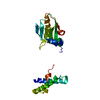
| ||||||||
|---|---|---|---|---|---|---|---|---|---|
| 1 |
| ||||||||
| Unit cell |
|
-Components
| #1: Protein | Mass: 25957.619 Da / Num. of mol.: 1 / Mutation: R38A, K41A Source method: isolated from a genetically manipulated source Source: (gene. exp.) Influenza A virus / Gene: NS1, NS / Production host:  Escherichia coli BL21(DE3) (bacteria) / Strain (production host): BL21(DE3) / References: UniProt: Q20NS3 Escherichia coli BL21(DE3) (bacteria) / Strain (production host): BL21(DE3) / References: UniProt: Q20NS3 |
|---|
-Experimental details
-Experiment
| Experiment | Method: X-RAY DIFFRACTION / Number of used crystals: 1 |
|---|
- Sample preparation
| Crystal | Density Matthews: 2.5 Å3/Da / Density % sol: 50.87 % |
|---|---|
| Crystal grow | Temperature: 293.15 K / Method: vapor diffusion, hanging drop / pH: 7 Details: 0.2 M Sodium citrate tribasic dehydrate, 0.1 M Tris hydrochloride pH 7.0, and 15% (v/v) PEG 400 |
-Data collection
| Diffraction | Mean temperature: 293.15 K / Serial crystal experiment: N |
|---|---|
| Diffraction source | Source: SYNCHROTRON / Site: ALS / Beamline: 8.2.1 / Wavelength: 0.999949 Å |
| Detector | Type: ADSC QUANTUM 315r / Detector: CCD / Date: Apr 13, 2016 |
| Radiation | Protocol: SINGLE WAVELENGTH / Monochromatic (M) / Laue (L): M / Scattering type: x-ray |
| Radiation wavelength | Wavelength: 0.999949 Å / Relative weight: 1 |
| Reflection | Resolution: 3.899→28.612 Å / Num. obs: 2536 / % possible obs: 95.2 % / Redundancy: 10.9 % / Rmerge(I) obs: 0.136 / Net I/σ(I): 12.8 |
| Reflection shell | Resolution: 3.899→4 Å / Rmerge(I) obs: 0.94 / Num. unique obs: 196 / % possible all: 99.4 |
- Processing
| Software |
| ||||||||||||||||||||||||||||||||||||||||
|---|---|---|---|---|---|---|---|---|---|---|---|---|---|---|---|---|---|---|---|---|---|---|---|---|---|---|---|---|---|---|---|---|---|---|---|---|---|---|---|---|---|
| Refinement | Method to determine structure: MOLECULAR REPLACEMENT Starting model: 4OPH Resolution: 3.899→28.612 Å / SU ML: 0.27 / Cross valid method: FREE R-VALUE / σ(F): 1.34 / Phase error: 23.36
| ||||||||||||||||||||||||||||||||||||||||
| Solvent computation | Shrinkage radii: 0.9 Å / VDW probe radii: 1.11 Å | ||||||||||||||||||||||||||||||||||||||||
| Refinement step | Cycle: LAST / Resolution: 3.899→28.612 Å
| ||||||||||||||||||||||||||||||||||||||||
| Refine LS restraints |
| ||||||||||||||||||||||||||||||||||||||||
| LS refinement shell | Resolution: 3.899→28.6131 Å
| ||||||||||||||||||||||||||||||||||||||||
| Refinement TLS params. | Method: refined / Origin x: 60.6795 Å / Origin y: 50.1299 Å / Origin z: -42.86 Å
| ||||||||||||||||||||||||||||||||||||||||
| Refinement TLS group | Selection details: all |
