 Movie
Movie Controller

[English] 日本語
 Yorodumi
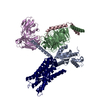
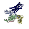
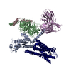
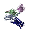
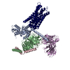
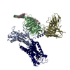
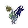
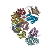
- PDB-6omm: Cryo-EM structure of formyl peptide receptor 2/lipoxin A4 recepto... -
+ Open data

- Basic information
| Entry | Database: PDB / ID: 6omm | |||||||||||||||
|---|---|---|---|---|---|---|---|---|---|---|---|---|---|---|---|---|
| Title | Cryo-EM structure of formyl peptide receptor 2/lipoxin A4 receptor in complex with Gi | |||||||||||||||
 Components Components |
| |||||||||||||||
 Keywords Keywords | SIGNALING PROTEIN / Formyl peptide receptor 2/lipoxin A4 receptor / GPCR / Gi protein | |||||||||||||||
| Function / homology |  Function and homology information Function and homology informationN-formyl peptide receptor activity / complement receptor activity / immune response-regulating cell surface receptor signaling pathway / scavenger receptor binding / RAGE receptor binding / complement receptor mediated signaling pathway / positive regulation of monocyte chemotaxis / positive regulation of innate immune response / Formyl peptide receptors bind formyl peptides and many other ligands / cargo receptor activity ...N-formyl peptide receptor activity / complement receptor activity / immune response-regulating cell surface receptor signaling pathway / scavenger receptor binding / RAGE receptor binding / complement receptor mediated signaling pathway / positive regulation of monocyte chemotaxis / positive regulation of innate immune response / Formyl peptide receptors bind formyl peptides and many other ligands / cargo receptor activity / positive chemotaxis / tertiary granule membrane / ficolin-1-rich granule membrane / adenylate cyclase inhibitor activity / specific granule membrane / positive regulation of protein localization to cell cortex / T cell migration / Adenylate cyclase inhibitory pathway / D2 dopamine receptor binding / response to prostaglandin E / positive regulation of phagocytosis / G protein-coupled serotonin receptor binding / adenylate cyclase regulator activity / adenylate cyclase-inhibiting serotonin receptor signaling pathway / cellular response to forskolin / receptor-mediated endocytosis / regulation of mitotic spindle organization / positive regulation of superoxide anion generation / astrocyte activation / Regulation of insulin secretion / microglial cell activation / positive regulation of cholesterol biosynthetic process / calcium-mediated signaling / G protein-coupled receptor binding / negative regulation of insulin secretion / G protein-coupled receptor activity / adenylate cyclase-inhibiting G protein-coupled receptor signaling pathway / response to peptide hormone / adenylate cyclase-modulating G protein-coupled receptor signaling pathway / negative regulation of inflammatory response / centriolar satellite / G-protein beta/gamma-subunit complex binding / Olfactory Signaling Pathway / Activation of the phototransduction cascade / G beta:gamma signalling through PLC beta / Presynaptic function of Kainate receptors / Thromboxane signalling through TP receptor / G protein-coupled acetylcholine receptor signaling pathway / G-protein activation / Activation of G protein gated Potassium channels / Inhibition of voltage gated Ca2+ channels via Gbeta/gamma subunits / Prostacyclin signalling through prostacyclin receptor / G beta:gamma signalling through CDC42 / Glucagon signaling in metabolic regulation / cellular response to amyloid-beta / chemotaxis / G beta:gamma signalling through BTK / Synthesis, secretion, and inactivation of Glucagon-like Peptide-1 (GLP-1) / ADP signalling through P2Y purinoceptor 12 / Sensory perception of sweet, bitter, and umami (glutamate) taste / photoreceptor disc membrane / Glucagon-type ligand receptors / Adrenaline,noradrenaline inhibits insulin secretion / Vasopressin regulates renal water homeostasis via Aquaporins / GDP binding / G alpha (z) signalling events / Glucagon-like Peptide-1 (GLP1) regulates insulin secretion / cellular response to catecholamine stimulus / ADP signalling through P2Y purinoceptor 1 / ADORA2B mediated anti-inflammatory cytokines production / G beta:gamma signalling through PI3Kgamma / Cooperation of PDCL (PhLP1) and TRiC/CCT in G-protein beta folding / adenylate cyclase-activating dopamine receptor signaling pathway / GPER1 signaling / Inactivation, recovery and regulation of the phototransduction cascade / cellular response to prostaglandin E stimulus / G-protein beta-subunit binding / heterotrimeric G-protein complex / G alpha (12/13) signalling events / sensory perception of taste / extracellular vesicle / signaling receptor activity / signaling receptor complex adaptor activity / Thrombin signalling through proteinase activated receptors (PARs) / amyloid-beta binding / retina development in camera-type eye / G protein activity / positive regulation of cytosolic calcium ion concentration / GTPase binding / Ca2+ pathway / midbody / fibroblast proliferation / High laminar flow shear stress activates signaling by PIEZO1 and PECAM1:CDH5:KDR in endothelial cells / cell cortex / G alpha (i) signalling events / G alpha (s) signalling events / phospholipase C-activating G protein-coupled receptor signaling pathway / Hydrolases; Acting on acid anhydrides; Acting on GTP to facilitate cellular and subcellular movement / G alpha (q) signalling events / negative regulation of neuron apoptotic process Similarity search - Function | |||||||||||||||
| Biological species |  Homo sapiens (human) Homo sapiens (human)synthetic construct (others) | |||||||||||||||
| Method | ELECTRON MICROSCOPY / single particle reconstruction / cryo EM / Resolution: 3.17 Å | |||||||||||||||
 Authors Authors | Zhuang, Y. / Liu, H. / de Waal, P.W. / Zhou, X.E. / Wang, L. / Meng, X. / Zhao, G. / Kang, Y. / Melcher, K. / Xu, H.E. / Zhang, C. | |||||||||||||||
| Funding support |  United States, 2items United States, 2items
| |||||||||||||||
 Citation Citation | Journal: Nat Commun / Year: 2020 Title: Structure of formylpeptide receptor 2-G complex reveals insights into ligand recognition and signaling. Authors: Youwen Zhuang / Heng Liu / X Edward Zhou / Ravi Kumar Verma / Parker W de Waal / Wonjo Jang / Ting-Hai Xu / Lei Wang / Xing Meng / Gongpu Zhao / Yanyong Kang / Karsten Melcher / Hao Fan / ...Authors: Youwen Zhuang / Heng Liu / X Edward Zhou / Ravi Kumar Verma / Parker W de Waal / Wonjo Jang / Ting-Hai Xu / Lei Wang / Xing Meng / Gongpu Zhao / Yanyong Kang / Karsten Melcher / Hao Fan / Nevin A Lambert / H Eric Xu / Cheng Zhang /   Abstract: Formylpeptide receptors (FPRs) as G protein-coupled receptors (GPCRs) can recognize formylpeptides derived from pathogens or host cells to function in host defense and cell clearance. In addition, ...Formylpeptide receptors (FPRs) as G protein-coupled receptors (GPCRs) can recognize formylpeptides derived from pathogens or host cells to function in host defense and cell clearance. In addition, FPRs, especially FPR2, can also recognize other ligands with a large chemical diversity generated at different stages of inflammation to either promote or resolve inflammation in order to maintain a balanced inflammatory response. The mechanism underlying promiscuous ligand recognition and activation of FPRs is not clear. Here we report a cryo-EM structure of FPR2-G signaling complex with a peptide agonist. The structure reveals a widely open extracellular region with an amphiphilic environment for ligand binding. Together with computational docking and simulation, the structure suggests a molecular basis for the recognition of formylpeptides and a potential mechanism of receptor activation, and reveals conserved and divergent features in G coupling. Our results provide a basis for understanding the molecular mechanism of the functional promiscuity of FPRs. | |||||||||||||||
| History |
|
- Structure visualization
| Movie |
Movie viewer |
|---|---|
| Structure viewer | Molecule: MolmilJmol/JSmol |
- Downloads & links
-Download
| PDBx/mmCIF format | 6omm.cif.gz | 247 KB | Display | PDBx/mmCIF format |
|---|---|---|---|---|
| PDB format | pdb6omm.ent.gz | 191.6 KB | Display | PDB format |
| PDBx/mmJSON format | 6omm.json.gz | Tree view | PDBx/mmJSON format | |
| Others |  Other downloads Other downloads |
-Validation report
| Summary document | 6omm_validation.pdf.gz | 1.2 MB | Display | wwPDB validaton report |
|---|---|---|---|---|
| Full document | 6omm_full_validation.pdf.gz | 1.2 MB | Display | |
| Data in XML | 6omm_validation.xml.gz | 36.6 KB | Display | |
| Data in CIF | 6omm_validation.cif.gz | 55.9 KB | Display | |
| Arichive directory | https://data.pdbj.org/pub/pdb/validation_reports/om/6ommftp://data.pdbj.org/pub/pdb/validation_reports/om/6omm | HTTPS FTP |
-Related structure data
| Related structure data | 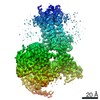 20126MC M: map data used to model this data C: citing same article ( |
|---|---|
| Similar structure data |

-Links
 PDBj
PDBj


- Assembly
| Deposited unit | 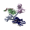
|
|---|---|
| 1 |
|
-Components
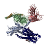
-Guanine nucleotide-binding protein ... , 3 types, 3 molecules ABC
| #3: Protein | Mass: 40327.891 Da / Num. of mol.: 1 / Mutation: G203A, A326S, D328E Source method: isolated from a genetically manipulated source Source: (gene. exp.) Homo sapiens (human) / Gene: GNAI1 / Production host:   Spodoptera frugiperda (fall armyworm) / References: UniProt: P63096 Spodoptera frugiperda (fall armyworm) / References: UniProt: P63096 |
|---|---|
| #4: Protein | Mass: 39021.648 Da / Num. of mol.: 1 / Mutation: Q6E, E130Q, N237D Source method: isolated from a genetically manipulated source Source: (gene. exp.) Homo sapiens (human) / Gene: GNB1 / Production host: Spodoptera frugiperda (fall armyworm) / References: UniProt: P62873 |
| #5: Protein | Mass: 7430.584 Da / Num. of mol.: 1 / Mutation: E17Q, E58Q Source method: isolated from a genetically manipulated source Source: (gene. exp.) Homo sapiens (human) / Gene: GNG2 / Production host: Spodoptera frugiperda (fall armyworm) / References: UniProt: P59768 |
-Protein / Protein/peptide / Antibody , 3 types, 3 molecules RLE
| #1: Protein | Mass: 40329.238 Da / Num. of mol.: 1 Source method: isolated from a genetically manipulated source Source: (gene. exp.) Homo sapiens (human) / Gene: FPR2, FPRH1, FPRL1, LXA4R / Production host: Spodoptera frugiperda (fall armyworm) / References: UniProt: P25090 |
|---|---|
| #2: Protein/peptide | Mass: 857.117 Da / Num. of mol.: 1 / Source method: obtained synthetically / Source: (synth.) synthetic construct (others) |
| #6: Antibody | Mass: 26323.324 Da / Num. of mol.: 1 Source method: isolated from a genetically manipulated source Source: (gene. exp.) synthetic construct (others) / Production host:  |
-Non-polymers , 2 types, 8 molecules 

| #7: Chemical | ChemComp-CLR /  Mass: 386.654 Da / Num. of mol.: 5 / Source method: obtained synthetically / Formula: C27H46O Mass: 386.654 Da / Num. of mol.: 5 / Source method: obtained synthetically / Formula: C27H46O#8: Chemical |  Mass: 256.424 Da / Num. of mol.: 3 / Source method: obtained synthetically / Formula: C16H32O2 Mass: 256.424 Da / Num. of mol.: 3 / Source method: obtained synthetically / Formula: C16H32O2 |
|---|
-Details
| Has ligand of interest | Y |
|---|---|
| Has protein modification | Y |
-Experimental details
-Experiment
| Experiment | Method: ELECTRON MICROSCOPY |
|---|---|
| EM experiment | Aggregation state: PARTICLE / 3D reconstruction method: single particle reconstruction |
- Sample preparation
| Component | Name: FPR2-Gi complex / Type: COMPLEX / Entity ID: #1-#6 / Source: RECOMBINANT |
|---|---|
| Source (natural) | Organism: Homo sapiens (human) |
| Source (recombinant) | Organism: Spodoptera frugiperda (fall armyworm) |
| Buffer solution | pH: 7.2 |
| Specimen | Embedding applied: NO / Shadowing applied: NO / Staining applied: NO / Vitrification applied: YES |
| Specimen support | Details: unspecified |
| Vitrification | Cryogen name: ETHANE |
- Electron microscopy imaging
| Experimental equipment |  Model: Titan Krios / Image courtesy: FEI Company |
|---|---|
| Microscopy | Model: FEI TITAN KRIOS |
| Electron gun | Electron source:  FIELD EMISSION GUN / Accelerating voltage: 300 kV / Illumination mode: FLOOD BEAM FIELD EMISSION GUN / Accelerating voltage: 300 kV / Illumination mode: FLOOD BEAM |
| Electron lens | Mode: BRIGHT FIELD |
| Image recording | Electron dose: 8.4 e/Å2 / Film or detector model: GATAN K2 BASE (4k x 4k) |
- Processing
| Software | Name: PHENIX / Version: 1.13_2998: / Classification: refinement |
|---|---|
| EM software | Name: PHENIX / Category: model refinement |
| CTF correction | Type: PHASE FLIPPING AND AMPLITUDE CORRECTION |
| 3D reconstruction | Resolution: 3.17 Å / Resolution method: FSC 0.143 CUT-OFF / Num. of particles: 1231594 / Symmetry type: POINT |
