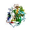
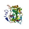
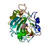
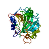
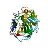
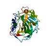
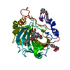
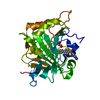
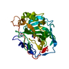
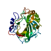
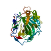
Entry Database : PDB / ID : 6oe1Title Benzensulfonamides bearing spyrohydantoin moieties act as potent inhibitors of human carbonic anhydrases II and VII and show neuropathic pain attenuating effects Carbonic anhydrase 2 Keywords / / / / Function / homology Function Domain/homology Component
/ / / / / / / / / / / / / / / / / / / / / / / / / / / / / / / / / / / / / / / / / / Biological species Homo sapiens (human)Method / / / Resolution : 1.45 Å Authors Peat, T.S. Journal : Eur.J.Med.Chem. / Year : 2019Title : Benzensulfonamides bearing spyrohydantoin moieties act as potent inhibitors of human carbonic anhydrases II and VII and show neuropathic pain attenuating effects.Authors : Angeli, A. / Di Cesare Mannelli, L. / Ghelardini, C. / Peat, T.S. / Bartolucci, G. / Menicatti, M. / Carta, F. / Supuran, C.T. History Deposition Mar 27, 2019 Deposition site / Processing site Revision 1.0 Jun 12, 2019 Provider / Type Revision 1.1 Oct 11, 2023 Group / Database references / Refinement descriptionCategory chem_comp_atom / chem_comp_bond ... chem_comp_atom / chem_comp_bond / database_2 / pdbx_initial_refinement_model Item / _database_2.pdbx_database_accession
Show all Show less
 Movie
Movie Controller

 Yorodumi
 Open data
 Basic information
 Components
 Keywords
 Function and homology information
 Homo sapiens (human)
 X-RAY DIFFRACTION /
  Authors
 Citation
 Structure visualization
 Downloads & links
 Other downloads
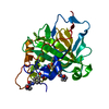


 PDBj
PDBj

 Assembly


 Mass: 65.409 Da / Num. of mol.: 1 / Source method: obtained synthetically / Formula: Zn
 Mass: 92.094 Da / Num. of mol.: 1 / Source method: obtained synthetically / Formula: C3H8O3
 Mass: 380.419 Da / Num. of mol.: 1 / Source method: obtained synthetically / Formula: C16H20N4O5S
 Mass: 96.063 Da / Num. of mol.: 1 / Source method: obtained synthetically / Formula: SO4
 Sample preparation
 / Beamline: MX1 / Wavelength: 0.9537 Å
 Processing